42 label the photomicrograph
A & P lab test 4 Flashcards | Quizlet Select all that apply. Label these structures of the upper respiratory system. Correctly label the components of the upper respiratory tract. Label the anterior view of the lower respiratory tract based on the hints if provided. Correctly label the components of the lungs. Correctly label the components of the pulmonary alveoli. Label The Photomicrograph Of Thin Skin. : Chapter 13, Page 3 ... Label The Photomicrograph Of Thin Skin. : Chapter 13, Page 3 - HistologyOLM Academia.edu is a platform for academics to share research papers. The beam is directed at a low angle to the specimen so that it acquires a "shadow" in the form of an uncoated area on the other side. 28.09.2020 · this involves depositing a thin layer of heavy metal ...
Ch. 22 Assessment Flashcards | Quizlet Label the structures in the photomicrograph based on the hints provided. List the correct order of lymphatic flow through a lymph node. 1. Afferent lymphatic vessel 2. Subcapsular sinus of the cortex 3. Sinuses of cortex and medulla 4. Efferent lymphatic vessel Put the following events into the correct order. 1.

Label the photomicrograph
Label the photomicrograph in figure 128 figure 128 - Course Hero Label the photomicrograph in Figure 12.8 Figure 12.8. LAB ACTIVITY 3: Neuromuscular Junction Examine a prepared microscope slide of the neuromuscular junction and identify the structures listed in Figure 12.8 Figure 12.8. Label The Photomicrograph Of Compact Bone. / Tik Ta Lk Aitee Umoffong ... Free answer to label the photomicrograph of compact bone. These bones do everything from protecting vital organs to giving muscles and nerves an anchor. (b) in this micrograph of the osteon, you can clearly see the concentric . The walls of the diaphysis are composed of dense and hard compact bone. 1 2 label the photomicrograph of a transverse section 1 2 Label the photomicrograph of a transverse section of the spinal cord in Figure 17.5 Figure 17.5. LAB ACTIVITY 3: Transverse Section of Spinal Cord Identify the spinal cord structures in Figures 17.3 Figures 17.3 and 17.4 17.4 on a transverse section model or chart of the spinal cord, or use the search text box in Real Anatomy Real Anatomy (Nervous) to find these structures.
Label the photomicrograph. Endocrine System APR Module 8 Flashcards - Quizlet Label the photomicrograph based on the hints provided. (suprarenal gland) Label each of the following histology slides by dragging the histology slide of the gland under the correct name. [Solved] Please see an attachment for details | Course Hero Label the photomicrograph based on the hints provided. Medulla Capillary Zona fasciculara Suprarenal gland Zona reticularis... Biology Science Anatomy BISC 106. ... Make sure you label every questions you answers so i could be easy to follow. organize is key Part A 1. Define anatomy a. Q: ... Label The Photomicrograph Based On The Hints Provided / Endocrine Lab ... Label The Photomicrograph Based On The Hints Provided / Endocrine Lab Flashcards Quizlet. Spleen capsule capsule white pulp. Can be reproduced based on the information provided in the manuscript. A study based on observation and interview with individuals that uses inductive. Medulla capillary zona fasciculara suprarenal gland zona reticularis. Final Exam A&P 1 Flashcards | Quizlet Label the photomicrograph of thin skin Hair shaft, epidermis, dermal root sheath, sebaceous gland, dermis, hair matrix label the structures of the hair follicle Identify the layers of the epidermis with relation to their location and role in keratinization ... the receptors responsible for olfaction are located in the olfactory epithelium
Anatomy and Physiology Homework Chapter 6 Flashcards - Quizlet Label the photomicrograph of thick skin.-Stratum corneum-Stratum granulosum-Stratum spinosum-Stratum basale-Epidermis-Dermis-Stratum lucidum-Epidermis-Stratum corneum-Stratum lucidum-Stratum granulosum-Stratum spinosum-Stratum basale-Dermis Explanation: Thick skin is located on the palms and soles. Refer to APR 3.0 for further information. Label The Photomicrograph Using The Hints Provided / The Fate And ... Label The Photomicrograph Using The Hints Provided / The Fate And Toxicity Of Raman Active Silica Gold Nanoparticles In Mice. Correctly label the following anatomical parts of a kidney. Correctly label the following … Contribute to cth/sdcg development by creating an account on github. Solved > Question 31 points Label the photomicrograph of ... - ScholarOn Question : Question 31 points Label the photomicrograph of thin skin. Hair Follicle : 391984. Question. 31 points Label the photomicrograph of thin skin. Hair Follicle Hair Dermis Sebaceous gland Duct of sebaceous gland Reset zoom. Solution. 5 (1 Ratings ) Solved. Biology 2 Years Ago 68 Views. Label The Photomicrograph Based On The Hints Provided. Capsule - 33 ... Label the photomicrograph based on the hints provided. Place each of the following lymphatic structures in the correct category based on their location. Searchable by topic and provided in ms word format, as well as in launchpad and diploma, the assessment bank offers a high level of flexibility.
Label The Photomicrograph Of The Sebaceous Gland : Histochemical ... 1 answer to label the photomicrograph of thin skin. If the gland become blocked, the sebum can be forced out into the dermis, where it elicits an inflammatory response. Photomicrograph of prepuce in golden jackal, (d) dermis, (sc) sebaceous gland, (sw) sweat gland, (g) guard hair follicle. fuiadinda64 April 01, 2022 URL Print Email Label The Photomicrograph Based On The Hints Provided. Zona Fasciculata ... Label the photomicrograph based on the hints provided. The cortex can be divided into three regions: Label the anterior view of the larynx based on the hints if provided. Adrenal gland · the zona glomerulosa is the thin outer layer of the adrenal cortex. Its cells are pale staining and organized in ovoid clusters that are separated by . Question: Label The Structures In The Photomicrograph Based On The ... Label the structures in the photomicrograph based on the hints provided. Mantle zone Lymph node Subcapsular sinus Germinal center Capsule Mwl Educationreg Reader (Visited 169 times, 1 visits today) Is this your assignment or some part of it? We can do it for you! Free Features. Answered: In the photomicrograph below of compact… | bartleby Transcribed Image Text: In the photomicrograph below of compact bone tissue, find and label the indicated structures Osteon Lamella Lacuna Osteocyte Canaliculi Central canal 1. Obtain a slide of ground compact bone connective tissue from the slide box. 2. View the slide on an appropriate objective. 3. Fill out the blanks next to your drawing. 4.
Photomicrograph of Plasmodium falciparum, the parasite that causes ...
Label the photomicrograph based on the hints provided. Pancreas ... Label the photomicrograph based on the hints provided. Pancreas Pancreatic islet Exocrine portion Pancreas Exocrine portion Intralobular duct Venule Pancreatic islet Arteriole Mar 29 2022 11:34 AM
Print Anatomy and Physiology 2 Chapter 17 flashcards | Easy Notecards
Label the photomicrograph based on the hints provided. the normal arterial oxygen is about 75 to 100 mm hg. the values underneath 60 mm hg is generally considered with the requirement for supplemental oxygen. the normal pulse oximeter readings generally range between 95 to 100 percent. the values under 90 percent are regarded as low.

Bio. Sci. 4 Microscopic Images
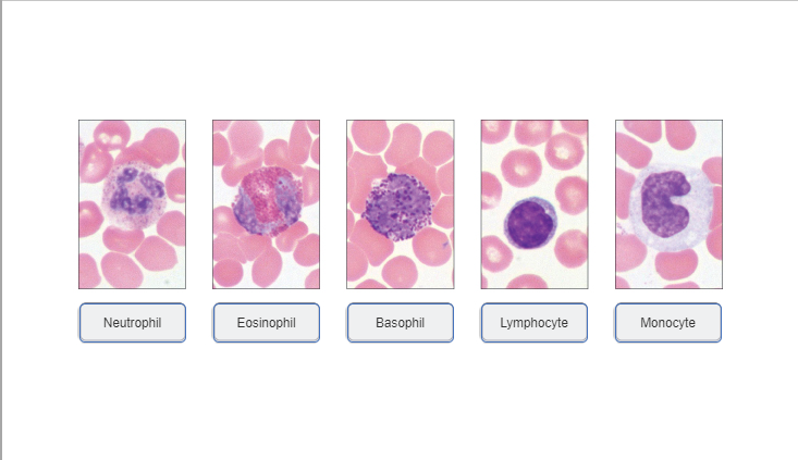
Label The Photomicrograph Using The Hints Provided : Pollen Microscope ... Photographic images can be taken through the microscope (photomicrography) by attaching a camera on to the vertical tube of the microscope's trinocular head label the photomicrograph. Using highly adherent human cervical tumor (hela) cells as a model. Routine stains are those used. Agranulocytes (includes lymphocytes and monocytes).

Integumentary System - Gland Development - Embryology
Label The Photomicrograph Of The Lung : 4 Chloro Dl Phenylalanine ... Label the photomicrogram of the lung segmental branch of pulmonary a. Make sure you know the basics of lung cancer, including prevention, risk factors, symptoms and treatment options. Label the anterior view of the lower respiratory tract based on the hints if. Electron micrograph of lung tissue (click to show / hide labels).

Zoeken in galerij
Solved Label the photomicrograph. Myoepithelial cell Lumen - Chegg We review their content and use your feedback to keep the quality high. 100% (13 ratings) The given micrograph is labelled and is attached below: Justification: Epithelial cell: The epithelial cells in mammary …. View the full answer. Transcribed image text: Label the photomicrograph. Myoepithelial cell Lumen Epithelial cell Apocrine sweat ...
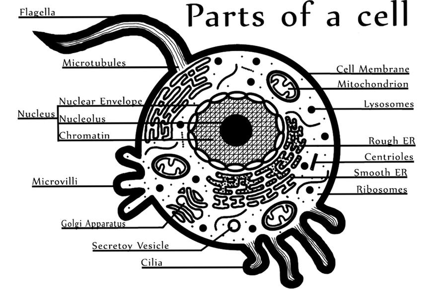
Diagram of an animal cell by andipoetic on DeviantArt
1 2 label the photomicrograph of a transverse section 1 2 Label the photomicrograph of a transverse section of the spinal cord in Figure 17.5 Figure 17.5. LAB ACTIVITY 3: Transverse Section of Spinal Cord Identify the spinal cord structures in Figures 17.3 Figures 17.3 and 17.4 17.4 on a transverse section model or chart of the spinal cord, or use the search text box in Real Anatomy Real Anatomy (Nervous) to find these structures.
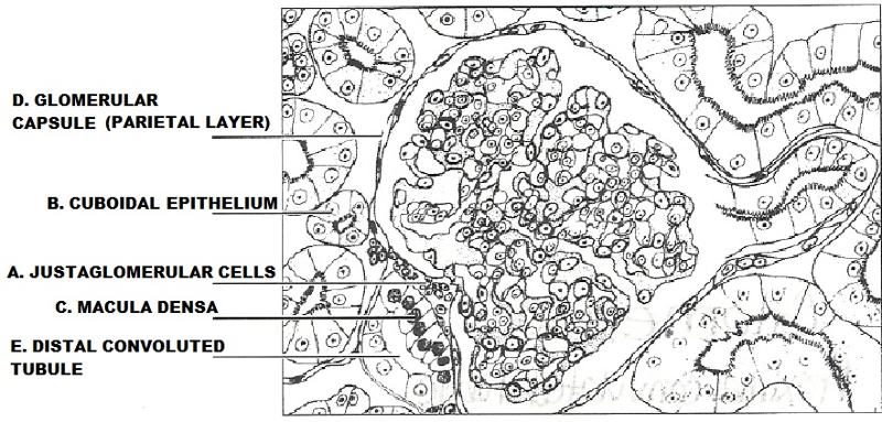
Exercise 40: Anatomy of the Urinary System Flashcards | Easy Notecards
Label The Photomicrograph Of Compact Bone. / Tik Ta Lk Aitee Umoffong ... Free answer to label the photomicrograph of compact bone. These bones do everything from protecting vital organs to giving muscles and nerves an anchor. (b) in this micrograph of the osteon, you can clearly see the concentric . The walls of the diaphysis are composed of dense and hard compact bone.

Photomicrograph of Thin Skin Quiz
Label the photomicrograph in figure 128 figure 128 - Course Hero Label the photomicrograph in Figure 12.8 Figure 12.8. LAB ACTIVITY 3: Neuromuscular Junction Examine a prepared microscope slide of the neuromuscular junction and identify the structures listed in Figure 12.8 Figure 12.8.

trachea histology labeled 400px respiratory histology 05 - Top Label Maker
Post a Comment for "42 label the photomicrograph"