43 label and color the parts of both microscopes
Compound Microscope Parts - Labeled Diagram and their Functions - Rs ... There are three major structural parts of a compound microscope. The head includes the upper part of the microscope, which houses the most critical optical components, and the eyepiece tube of the microscope. The base acts as the foundation of microscopes and houses the illuminator. The arm connects between the base and the head parts. Microscope Parts and Functions All of the parts of a microscope work together - The light from the illuminator passes through the aperture, through the slide, and through the objective lens, where the image of the specimen is magnified.
Answered: A. Draw the Diagram of Light Microscope… | bartleby Human Anatomy & Physiology (11th Edition) 11th Edition. ISBN: 9780134580999. Author: Elaine N. Marieb, Katja N. Hoehn. Publisher: PEARSON. expand_less. 1 The Human Body: An Orientation 2 Chemistry Comes Alive 3 Cells: The Living Units 4 Tissue: The Living Fabric 5 The Integumentary System 6 Bones And Skeletal Tissues 7 The Skeleton 8 Joints 9 ...

Label and color the parts of both microscopes
Parts of a Compound Microscope (And their Functions) Usually you'll get some yellow, green and blue filters to place in your condenser to normalize the light in case the color of the light doesn't feel natural. 10. Light Intensity Regulator (Dimmer) On the bottom right-hand side of the microscope (usually right by the base), you will find a light intensity regulator. Labeling the Parts of the Microscope Labeling the Parts of the Microscope. This activity has been designed for use in homes and schools. Each microscope layout (both blank and the version with answers) are available as PDF downloads. You can view a more in-depth review of each part of the microscope here. Download the Label the Parts of the Microscope PDF printable version here. Download the Label the Parts of the Microscope: Answers PDF printable version here. Color the Parts of the Microscope - The Biology Corner The arm is the correct place to grip the microscope when carrying it while supporting the base with the palm of your other hand. Color the arm green and the base red . The body tube (C) allows the light to pass upward to where the user's eye is. A lens inside the eyepiece (A) usually has a magnification of 10x.
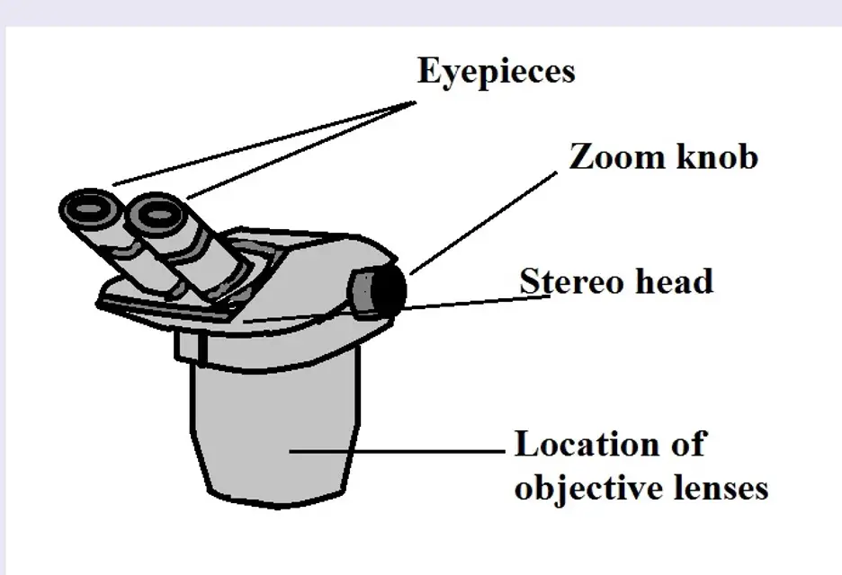
Label and color the parts of both microscopes. Parts of the Microscope with Labeling (also Free Printouts) Parts of Stereo Microscope (Dissecting microscope) - Rs' Science Optical parts of a stereo microscope work together to magnify and produce a 3-D image of the specimens. These parts include: Eyepieces. The eyepiece (or ocular lens) is the lens part at the top of a microscope that the viewer looks through. Typically, standard eyepieces for a dissecting microscope have a magnifying power of 10x. Parts of a microscope with functions and labeled diagram Apr 19, 2022 · Q. List down the 18 parts of a Microscope. 1. Ocular Lens (Eye Piece) 2. Diopter Adjustment 3. Head 4. Nose Piece 5. Objective Lens 6. Arm (Carrying Handle) 7. Mechanical Stage 8. Stage Clip 9. Aperture 10. Diaphragm 11. Condenser 12. Coarse Adjustment 13. Fine Adjustment 14. Illuminator (Light Source) 15. Stage Controls 16. Base 17. Brightness Adjustment 18. Light Switch PDF Label parts of the Microscope: Answers Label parts of the Microscope: Answers Coarse Focus Fine Focus Eyepiece Arm Rack Stop Stage Clip . Created Date: 20150715115425Z ...
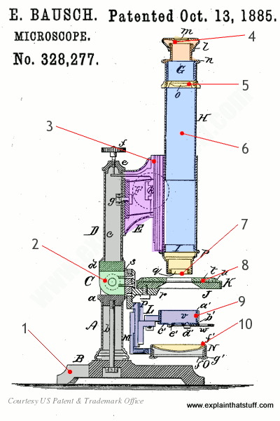
DOC Parts of a Microscope - coralgablescavaliers.org Always begin focusing a microscope on the lowest power and then move to the next higher power and refocus. Label and color. the low power objective pink and the high power objective red. The eyepiece is at the top of the body tube. Label. the body tube. The objective lenses are located on a . revolving nosepiece at the bottom of the body tube ... PDF Parts of a Microscope - Class Website Always begin focusing a microscope on the lowest power and then move to the next higher power and refocus. Label and color the low power objective pink and the high power objective red. The eyepiece is at the top of the body tube. Label the body tube. The objective lenses are located on a revolving nosepiece at the bottom of the body tube. A Study of the Microscope and its Functions With a Labeled Diagram To better understand the structure and function of a microscope, we need to take a look at the labeled microscope diagrams of the compound and electron microscope. These diagrams clearly explain the functioning of the microscopes along with their respective parts. Man's curiosity has led to great inventions. The microscope is one of them. Microscope Parts & Functions - AmScope Stereo Microscope: A low power microscope or dissecting microscope with a separate eyepiece and objective lens for each eye. These separate optical channels enable stereo or three-dimensional images of the specimen. See Compound Microscope. Sub-Stage: The parts of the microscope below the stage, including the illumination system.
Microscope, Microscope Parts, Labeled Diagram, and Functions Revolving Nosepiece or Turret: Turret is the part of the microscope that holds two or multiple objective lenses and helps to rotate objective lenses and also helps to easily change power. Objective Lenses: Three are 3 or 4 objective lenses on a microscope. The objective lenses almost always consist of 4x, 10x, 40x and 100x powers. The most common eyepiece lens is 10x and when it coupled with ... Label the microscope - Science Learning Hub Jun 08, 2018 · All microscopes share features in common. In this interactive, you can label the different parts of a microscope. Use this with the Microscope parts activity to help students identify and label the main parts of a microscope and then describe their functions. Drag and drop the text labels onto the microscope diagram. If you want to redo an answer, click on the box and the answer will go back to the top so you can move it to another box. DOC Parts of a Microscope - THOMAS Label and Color the Parts of both microscopes! Questions: 1. What is the difference between ocular and objective lenses? 2. What part of a microscope helps adjust the brightness of an image? 3. How should a microscope be carried? 4. The ocular and objectives are found at the top and bottom of what part of a microscope? 5. Microscope Diagram Labeled, Unlabeled and Blank | Parts of a Microscope Description A collection of microscope diagrams and worksheets for science class. Download them all in one convenient PDF, and select the version that's best for your classroom. This PDF contains the following: 1. Parts of a Microscope Diagram - Color 2. Parts of a Microscope Diagram - Black and White 3.

BW OPTICS
Compound Microscope: Parts of Compound Microscope - BYJUS (A) Mechanical Parts of a Compound Microscope 1. Foot or base It is a U-shaped structure and supports the entire weight of the compound microscope. 2. Pillar It is a vertical projection. This stands by resting on the base and supports the stage. 3. Arm The entire microscope is handled by a strong and curved structure known as the arm. 4. Stage
label and color the parts of both microscopes - Labels 2021
DOC Parts of a Microscope - Biology Junction Always begin focusing a microscope on the lowest power and then move to the next higher power and refocus. Label and color the low power objective pink and the high power objective red. The eyepiece is at the top of the body tube. Label the body tube. The objective lenses are located on a revolving nosepiece at the bottom of the body tube.
How does a microscope work? - Explain that Stuff
Microscope Parts | Microbus Microscope Educational Website Lenses are color coded and if built to DIN standards are interchangeable between microscopes. The high power objective lenses are retractable (ie 40xr). This means that if they hit a slide, the end of the lens will push in (spring loaded) thereby protecting the lens and the slide. All quality microscopes have achromatic, parcentered, parfocal ...

Labeling the Parts of the Microscope | Microscope activity, Apologia ...
PDF Parts of a Microscope reading assignment - White Plains Public Schools Label and color the low power objective pink and the high power objective red. The eyepiece is at the top of the body tube. Label the body tube. The objective lenses are located on a revolving nosepiece at the bottom of the body tube. Label and color the nosepiece brown and the body tube orange. 6. How should you always begin focusing? 7.

32 Label And Color The Parts Of Both Microscopes Answers - Labels ...
Parts of the Microscope Printables - ThoughtCo Coloring Page Beverly Hernandez Use this microscope coloring page just for fun or to occupy younger students while older siblings learn about and use their microscopes. Even young children will enjoy looking at specimens under a microscope, so invite your little ones to make observations, too. Theme Paper Beverly Hernandez

32 Label And Color The Parts Of Both Microscopes Answers - Labels For You
Light Microscope- Definition, Principle, Types, Parts, Labeled Diagram ... A light microscope is a biology laboratory instrument or tool, that uses visible light to detect and magnify very small objects and enlarge them. They use lenses to focus light on the specimen, magnifying it thus producing an image. The specimen is normally placed close to the microscopic lens.

Travel Inside 3D Cells in Full Color on Your Laptop
Exercise A 1 Label the parts of this stylized microscope... View Homework Help - Lab 2 Microscope Worksheet from BIO 103 at Paonia High School. 1 Worksheet for Microscope Lab week 2 Exercise A 1. Label the parts of this stylized microscope. Write the name of
Post a Comment for "43 label and color the parts of both microscopes"