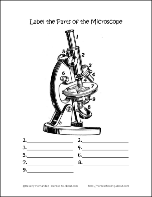
44 draw and label the compound microscope
How to draw compound of Microscope easily - step by step I will show you " How to draw compound of microscope easily - step by step "Please watch carefully and try this okay.Thanks for watching.....#microscopedrawi... Compound Microscope Parts, Function, & Diagram | What is a Compound ... The compound microscope, also called compound light microscope, is an upright microscope that utilizes two lenses to magnify objects. It gets its name because it uses two lenses added to each ...
Compound Microscope - Types, Parts, Diagram, Functions and Uses A compound microscope captures an inverted image of the specimen because every time the light passes through the lens, the image's direction is flipped. The image always ends up inverted from the original. So, if you move the sample to the left, it moves in the right direction. Image 18: A comparison image between a simple and compound microscope.

Draw and label the compound microscope
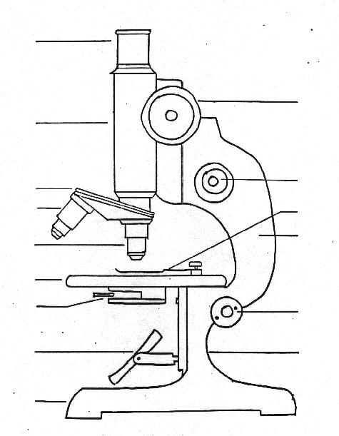
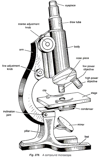
Microscope Drawing And Label - Painting Valley All the best Microscope Drawing And Label 33+ collected on this page. Feel free to explore, study and enjoy paintings with PaintingValley.com ... Compound Microscope ... 496x600 35 0. Like JPG. Parts Of A Compound ... 500x469 27 0. Like JPG. Microscopic Drawing ... 1024x1024 21 4. ... Draw A Large Diagram... 960x720 1 0. Like JPG. Drawings ... The Compound Microscope.docx - The Compound Microscope 1. Draw and ... Draw and label a compound microscope. Parts FunctionsParts of a Compound Microscope 2. Enumerate all the parts of the microscope and give their corresponding functions. Tabulate your answer. a. Objective Lenses -forms the inverted image of the specimen and gives the initial magnification. -used for visualization of specimen. c. Labelled Diagram of Compound Microscope - Biology Discussion The below mentioned article provides a labelled diagram of compound microscope. Part # 1. The Stand: The stand is made up of a heavy foot which carries a curved inclinable limb or arm bearing the body tube. The foot is generally horse shoe-shaped structure (Fig. 2) which rests on table top or any other surface on which the microscope in kept.
Draw and label the compound microscope. Label the microscope — Science Learning Hub Use this with the Microscope parts activity to help students identify and label the main parts of a microscope and then describe their functions. Drag and drop the text labels onto the microscope diagram. If you want to redo an answer, click on the box and the answer will go back to the top so you can move it to another box. (b) Why both objective and eyepiece of a compound microscope must have ... Question (a) Draw the labelled ray diagram for the formation of image by a compound microscope. Derive an expression for its total magnification (or magnifying power), when the final image is formed at the near point. (b) Why both objective and eyepiece of a compound microscope must have short focal lengths? how to draw microscope (compound) - YouTube drawing microscope. Thank you watching more videos.please subscribe my channel Working Principle and Parts of a Compound Microscope (with Diagrams) Therefore, the smallest details that can be seen by a typical light microscope is having the dimension of approximately 0.2 µ. Smaller objects or finer details than this cannot be resolved in a compound microscope. 5. Eyepiece: The eyepiece is a drum, which fits loosely into the draw tube.
Diagram of a Compound Microscope - Biology Discussion Magnification of the Image of the Object by Compound Microscope: A bright-field or compound microscope is primarily used to enlarge or magnify the image of the object that is being viewed, which can not otherwise be seen by the naked eye. Magnification may be defined as the degree of enlargement of the image of an object provided by the microscope. BIOL1100_Act 2 The Compound Microscope- Laboratory Report.docx BIOL 1100 Activity No. 2 The Compound Microscope 3. Sketch the letter "e" at each resolution and compute for total magnification (TM) Letter "e" @ 4X TM = Letter "e" @ 10X TM = Letter "e" @ 40X TM = Letter "e" @ 100X TM= Letter "e" viewed using a magnifying glass BIOL 1100 Activity No. 2 The Compound Microscope Answers to Questions: 1. Compound Microscope Parts - Labeled Diagram and their Functions - Rs ... The term "compound" refers to the microscope having more than one lens. Basically, compound microscopes generate magnified images through an aligned pair of the objective lens and the ocular lens. In contrast, "simple microscopes" have only one convex lens and function more like glass magnifiers. Compound Microscope Labeled Diagram | Quizlet QUESTION. The total magnification of a specimen being viewed with a 10X ocular lens and a 40X objective lens is. 15 answers. QUESTION. a mosquito beats its wings up and down 600 times per second, which you hear as a very annoying 600 Hz sound. if the air outside is 20 C, how far would a sound wave travel between wing beats. 2 answers.
draw and label the compound microscope - Brainly.ph Draw and label the compound microscope - 9474237 samanthasolito19 samanthasolito19 19.01.2021 Science Elementary School answered Draw and label the compound microscope 1 See answer Advertisement Advertisement gemjem60 gemjem60 Answer: here I hope this will help. Microscope Parts and Functions First, the purpose of a microscope is to magnify a small object or to magnify the fine details of a larger object in order to examine minute specimens that cannot be seen by the naked eye. Here are the important compound microscope parts... Eyepiece: The lens the viewer looks through to see the specimen. Draw a labelled diagram of an image formed by a compound microscope ... Draw a labelled diagram of an image formed by a compound microscope, with the image at least distance of distinct vision. Write any one expression for its magnifying power. Medium Solution Verified by Toppr Expression of magnifying power of a compound microscope is given by: m=− u ov o(1+ f eD) Parts of a microscope with functions and labeled diagram Head - This is also known as the body. It carries the optical parts in the upper part of the microscope. Base - It acts as microscopes support. It also carries microscopic illuminators. Arms - This is the part connecting the base and to the head and the eyepiece tube to the base of the microscope.
how to draw microscope step by step slow and medium speed - YouTube
Compound Microscope- Definition, Labeled Diagram, Principle, Parts, Uses A compound microscope is of great use in pathology labs so as to identify diseases. Various crime cases are detected and solved by drawing out human cells and examining them under the microscope in forensic laboratories. The presence or absence of minerals and the presence of metals can be identified using compound microscopes.
Microscope Drawing And Label at GetDrawings | Free download
Compound Microscope Parts, Functions, and Labeled Diagram The total magnification of a compound microscope is calculated by multiplying the objective lens magnification by the eyepiece magnification level. So, a compound microscope with a 10x eyepiece magnification looking through the 40x objective lens has a total magnification of 400x (10 x 40).

Biology%20190 a%20lab%201%20%20intro
Parts of a Compound Microscope (And their Functions) Parts of a Compound Microscope (And their Functions) By Chris Ramsay A compound microscope is the most common microscope you can get and the type you'll typically see in a lab or hobbyist's study. These microscopes tend to have total magnification between 40x - 2000x to allow you to see specimens like bacteria and cells.
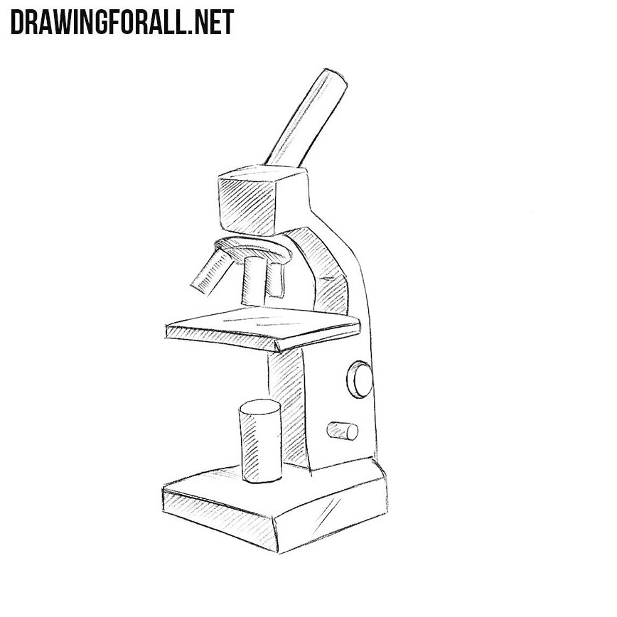
How to Draw a Microscope Easy
Compound Microscope: Definition, Diagram, Parts, Uses, Working ... - BYJUS A compound microscope is defined as A microscope with a high resolution and uses two sets of lenses providing a 2-dimensional image of the sample. The term compound refers to the usage of more than one lens in the microscope. Also, the compound microscope is one of the types of optical microscopes.

Light Microscope Sketch at PaintingValley.com | Explore collection of ...
Compound Microscope: Parts of Compound Microscope - BYJUS (A) Mechanical Parts of a Compound Microscope 1. Foot or base It is a U-shaped structure and supports the entire weight of the compound microscope. 2. Pillar It is a vertical projection. This stands by resting on the base and supports the stage. 3. Arm The entire microscope is handled by a strong and curved structure known as the arm. 4. Stage
Free Microscope Drawing, Download Free Microscope Drawing png images ...
Draw and label a compound microscope? - Answers Draw and label a compound microscope? Wiki User. ∙ 2012-09-19 09:42:29. Study now. See answer (1) Best Answer. Copy. uhmm,.... unang ana is the. 1.) Eyepiece. 2.) arm. eewan ko nakalimutan ko na ...
Light Microscope Drawing at GetDrawings | Free download
16 Parts of a Compound Microscope: Diagrams and Video In compound microscopes with two eye pieces there are prisms contained in the body that will also split the beam of light to enable you to view the image through both eye pieces. 2. Arm The arm of the microscope is another structural piece. The arm connects the base of the microscope to the head/body of the microscope.
Compound Microscope Drawing With Label - Micropedia
Draw a neat labelled diagram of a compound microscope and explain its ... Dividing and multiplying by I1 G1 on the right side, we get Magnifying power of the objective (m0) = I1G1/OJ = Height of the image due to the objective. Magnifying power of the eye piece (me) = IG/I1G1 = Height of the final image / Height of the object for the eyepiece. ∴ m = m0 × me ..... (1)

Microscope Clip Art at Clker.com - vector clip art online, royalty free ...
Compound Microscope - Diagram (Parts labelled), Principle and Uses See: Labeled Diagram showing differences between compound and simple microscope parts Structural Components The three structural components include 1. Head This is the upper part of the microscope that houses the optical parts 2. Arm This part connects the head with the base and provides stability to the microscope.
Post a Comment for "44 draw and label the compound microscope"