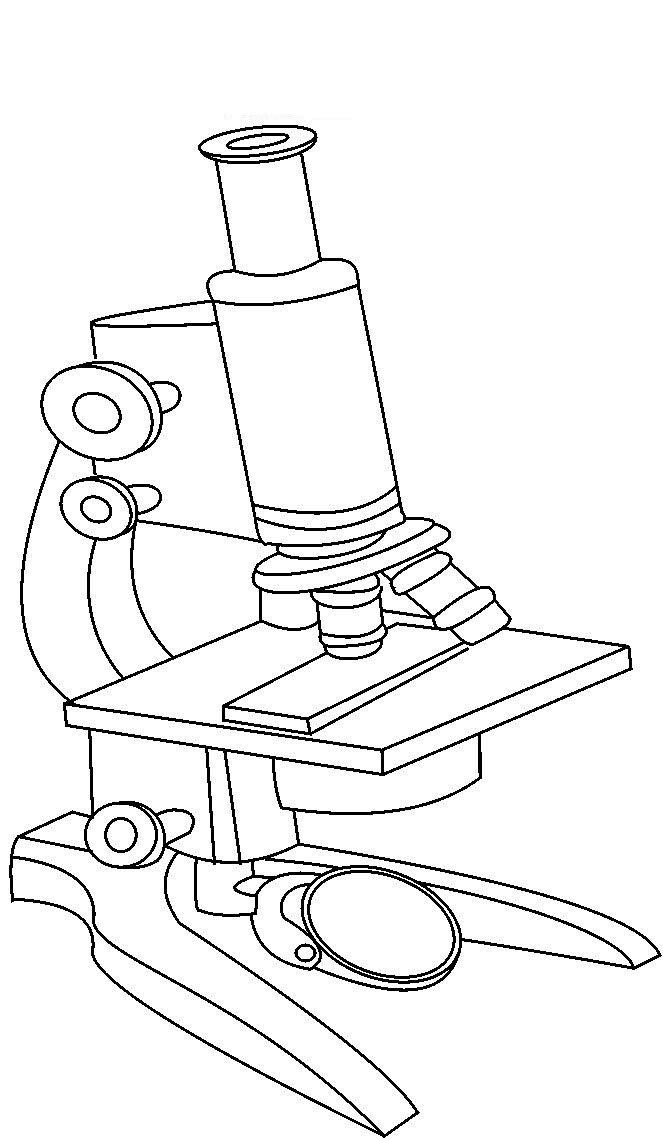
44 compound light microscope diagram labeled
Label the microscope - Science Learning Hub Label the microscope Add to collection Use this interactive to identify and label the main parts of a microscope. Drag and drop the text labels onto the microscope diagram. eye piece lens coarse focus adjustment high-power objective diaphragm or iris base fine focus adjustment light source stage Download Exercise Tweet Compound Microscope Parts - Labeled Diagram and their Functions - Rs ... Labeled diagram of a compound microscope Major structural parts of a compound microscope There are three major structural parts of a compound microscope. The head includes the upper part of the microscope, which houses the most critical optical components, and the eyepiece tube of the microscope.
Compound Light Microscope Drawing - Painting Valley All the best Compound Light Microscope Drawing 36+ collected on this page. Feel free to explore, study and enjoy paintings with PaintingValley.com. ... Labeled Diagram Of A... 300x181 0 0. Like JPG. Labeled Diagram Of C... 472x653 0 0. Like JPG. Light Microscopy Dra... 638x826 0 0. Like JPG. Microscope - Compoun... 236x305 0 0.

Compound light microscope diagram labeled
Parts of a Compound Microscope and Their Functions Compound microscope mechanical parts (Microscope Diagram: 2) include base or foot, pillar, arm, inclination joint, stage, clips, diaphragm, body tube, nose piece, coarse adjustment knob and fine adjustment knob.. Base: It's the horseshoe-shaped base structure of microscope.All of the other components of the compound microscope are supported by it. ... Ternary Phase Diagram - an overview | ScienceDirect Topics A point on the diagram represents a composition that is specified in terms of mole fraction or weight fraction. The point, (0.3, 0.4, 0.3) is at the center of the small triangle in the diagram and is located by following the red diagonal 60° line at red 0.3 and the horizontal line at blue 0.4 or any combination of two of the coordinates (A, B, C). PDF Parts of the Light Microscope - Science Spot Supports the MICROSCOPE D. STAGE CLIPS HOLD the slide in place C. OBJECTIVE LENSES Magnification ranges from 10 X to 40 X F. LIGHT SOURCE Projects light UPWARDS through the diaphragm, the SPECIMEN, and the LENSES H. DIAPHRAGM Regulates the amount of LIGHT on the specimen E. STAGE Supports the SLIDE being viewed K. ARM Used to SUPPORT the
Compound light microscope diagram labeled. Labeling the Parts of the Microscope | Microscope activity, Science ... Description Worksheet identifying the parts of the compound light microscope. Answer key: 1. Body tube 2. Revolving nosepiece 3. Low power objective 4. Medium power objective 5. High power objective 6. Stage clips 7. ... Print a microscope diagram, microscope worksheet, or practice microscope quiz in order to learn all the parts of a microscope ... Compound Microscope - Diagram (Parts labelled), Principle and Uses Compound Microscope - Diagram (Parts labelled), Principle and Uses As the name suggests, a compound microscope uses a combination of lenses coupled with an artificial light source to magnify an object at various zoom levels to study the object. A compound microscope: Is used to view samples that are not visible to the naked eye Compound Microscope Labeled Diagram | Quizlet QUESTION. The total magnification of a specimen being viewed with a 10X ocular lens and a 40X objective lens is. 15 answers. QUESTION. a mosquito beats its wings up and down 600 times per second, which you hear as a very annoying 600 Hz sound. if the air outside is 20 C, how far would a sound wave travel between wing beats. 2 answers. 13 parts of the Compound Light Microscope Diagram | Quizlet 13 parts of the Compound Light Microscope STUDY Learn Flashcards Write Spell Test PLAY Match Gravity Created by SchneiderFamilyStudy Terms in this set (15) base holds up microscope mirror directs ambient light up through the hole in the stage and illuminates specimen microscope stage flat platform that supports the stage microscope arm
Compound Light Microscope Diagram Worksheet - Google Groups Modern compound light microscopes under optimal conditions can we an average from 1000X to 2000X times the specimens original diameter Diagram. Label the parts of the microscope using the word... Compound Microscope Parts, Functions, and Labeled Diagram Compound Microscope Parts, Functions, and Labeled Diagram Parts of a Compound Microscope Each part of the compound microscope serves its own unique function, with each being important to the function of the scope as a whole. Simple Microscope- Definition, Principle, Magnification, Parts ... 06/05/2022 · A simple microscope is one that uses a single lens for magnification, such as a magnifying glass while a compound microscope uses several lenses to enhance the magnification of an object. ... Parts of a microscope with functions and labeled diagram; Light Microscope- Definition, Principle, Types, Parts, Labeled Diagram, Magnification; Microscope- Definition, Parts, Functions, Types, Diagram, Uses 21/02/2022 · A microscopic illuminator is a light source. In some compound microscope, the mirror is used which reflect the light from an external source to the sample. In other optical microscopes, different electric bulbs of low voltages are used as a constant light source. ... Types of Plant Cell- Definition, Structure, Functions, Labeled Diagram; Phylum ...
Parts of a microscope with functions and labeled diagram Figure: Diagram of parts of a microscope There are three structural parts of the microscope i.e. head, base, and arm. Head - This is also known as the body. It carries the optical parts in the upper part of the microscope. Base - It acts as microscopes support. It also carries microscopic illuminators. Two-Photon Excitation Microscopy for the Study of Living Cells … The development of miniature two-photon microscope systems and endoscopic or in vivo light delivery has broadened the range of sites than can be accessed. With such miniaturization, a two-photon microscope system can be mounted on freely moving mice, allowing longitudinal imaging studies ( Flusberg et al., 2005 ; Piyawattanametha et al., 2009 ). Solved Label the image of a compound light microscope using - Chegg Step-by-step answer. Who are the experts? Experts are tested by Chegg as specialists in their subject area. We review their content and use your feedback to keep the quality high. Transcribed image text: Label the image of a compound light microscope using the terms provided. Parts of Stereo Microscope (Dissecting microscope) – labeled diagram ... If you would like to learn optical components of a compound microscope, please visit Compound Microscope Parts – Labeled Diagram and their Functions, and this article. How to use a stereo (dissecting) microscope. Follow these steps to put your stereo microscopes in work: 1. Set your microscope on a tabletop or other flat sturdy surface where ...
Compound Microscope Parts, Function, & Diagram | What is a Compound ... Compound light microscopes are used to view microorganisms such as bacteria Compound light microscopes are used any time a scientist needs to view something that is too small to be seen with the...
Labelled Diagram of Compound Microscope - Biology Discussion The below mentioned article provides a labelled diagram of compound microscope. Part # 1. The Stand: The stand is made up of a heavy foot which carries a curved inclinable limb or arm bearing the body tube. The foot is generally horse shoe-shaped structure (Fig. 2) which rests on table top or any other surface on which the microscope in kept.
PDF COMPOUND LIGHT MICROSCOPE LAB - Springfield Public Schools COMPOUND(LIGHT(MICROSCOPE(LAB((Follow(written(andoral(instructions.((((A.((Label(the(parts(of(the(compound(microscope.(Be(able(to(label(a(blank(diagram
Light microscopes - Cell structure - Edexcel - BBC Bitesize The components of a light microscope and their functions Calculating the magnification of light microscopes. The compound microscope uses two lenses to magnify the specimen: the eyepiece and an ...
STED microscopy - Wikipedia STED microscopy is one of several types of super resolution microscopy techniques that have recently been developed to bypass the diffraction limit of light microscopy to increase resolution. STED is a deterministic functional technique that exploits the non-linear response of fluorophores commonly used to label biological samples in order to ...
PDF The Compound Light Microscope The Compound Light Microscope TASK Refer to page 605 in your text to: 1. Name each of the structures described in the table to the right. 2. Match each structure to the letter in the diagram below. ** ALWAYS USE TWO HANDS TO CARRY A MICROSCOPE** Letter Structure Function joins body tube to base supports the entire microscope
Compound Light Microscope: Everything You Need to Know A compound light microscope is a type of light microscope that uses a compound lens system, meaning, it operates through two sets of lenses to magnify the image of a specimen. It's an upright microscope that produces a two-dimensional image and has a higher magnification than a stereoscopic microscope. It also goes by a couple of other names ...
Pin on Science worksheets - Pinterest A Study of the Microscope and its Functions With a Labeled Diagram To better understand the structure and function of a microscope, we need to take a look at the labeled microscope diagrams of the compound and electron microscope. These diagrams clearly explain the functioning of the microscopes along with their respective parts. M mooketsi
Compound Microscope: Definition, Diagram, Parts, Uses, Working ... - BYJUS The parts of a compound microscope can be classified into two: Non-optical parts Optical parts Non-optical parts Base The base is also known as the foot which is either U or horseshoe-shaped. It is a metallic structure that supports the entire microscope. Pillar The connection between the base and the arm are possible through the pillar. Arm


Post a Comment for "44 compound light microscope diagram labeled"