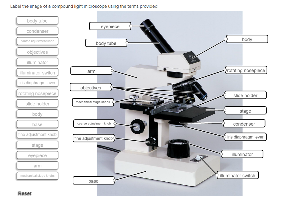
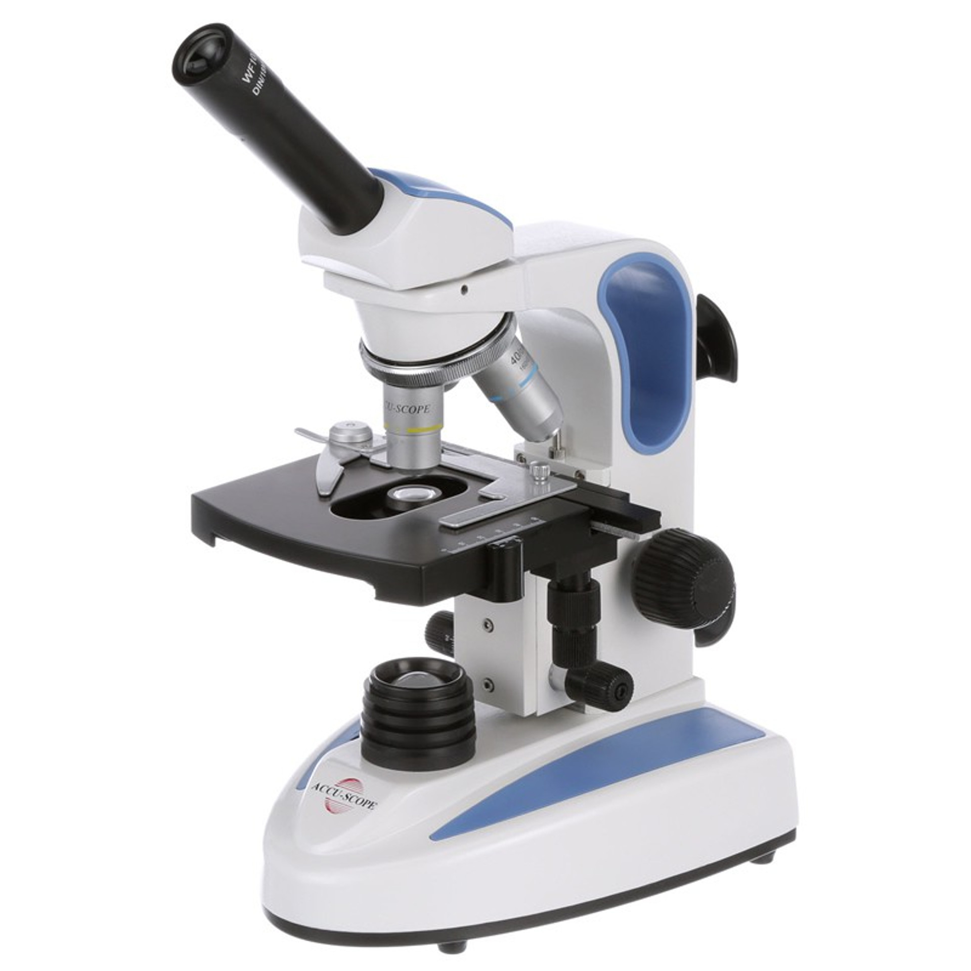
39 label a compound light microscope
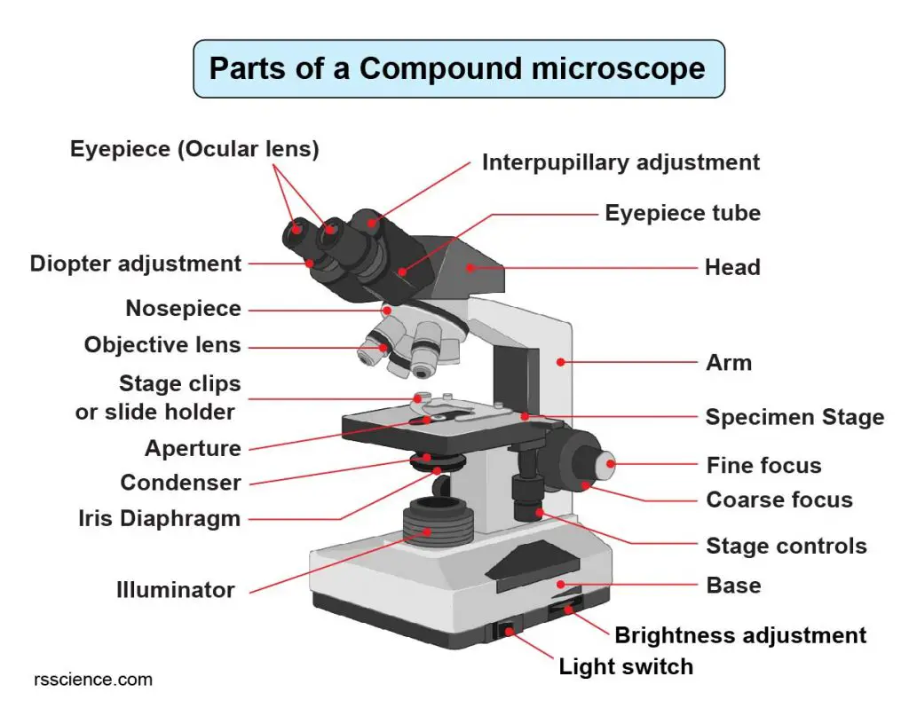
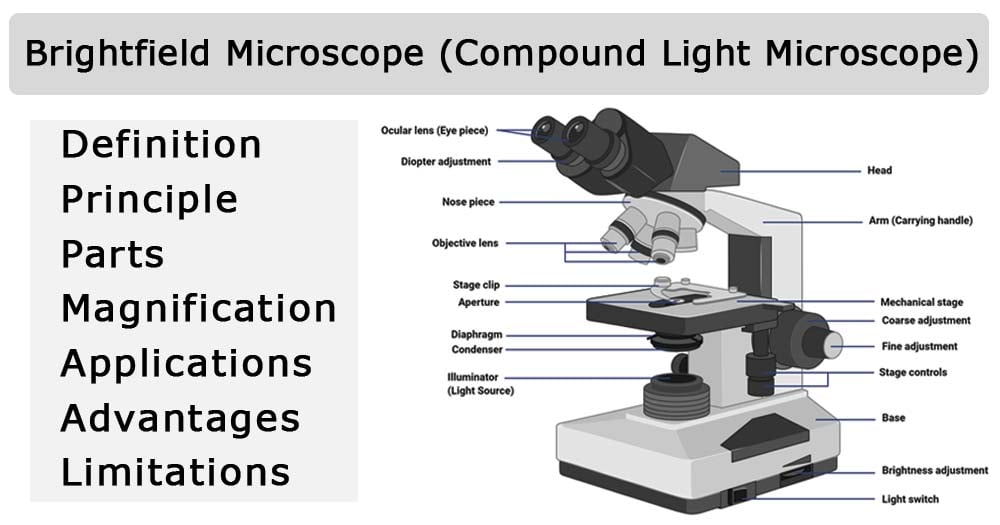
Microscope Parts and Functions First, the purpose of a microscope is to magnify a small object or to magnify the fine details of a larger object in order to examine minute specimens that cannot be seen by the naked eye. Here are the important compound microscope parts... Eyepiece: The lens the viewer looks through to see the specimen. Labelled Diagram of Compound Microscope - Biology Discussion The below mentioned article provides a labelled diagram of compound microscope. Part # 1. The Stand: The stand is made up of a heavy foot which carries a curved inclinable limb or arm bearing the body tube. The foot is generally horse shoe-shaped structure (Fig. 2) which rests on table top or any other surface on which the microscope in kept.
Compound Microscope- Definition, Labeled Diagram, Principle, Parts, Uses Alternatively, the magnification of the compound microscope is given by: m = D/ fo * L/fe where, D = Least distance of distinct vision (25 cm) L = Length of the microscope tube fo = Focal length of the objective lens fe = Focal length of the eye-piece lens Parts of a Compound Microscope Eyepiece And Body Tube.

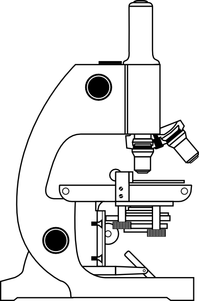
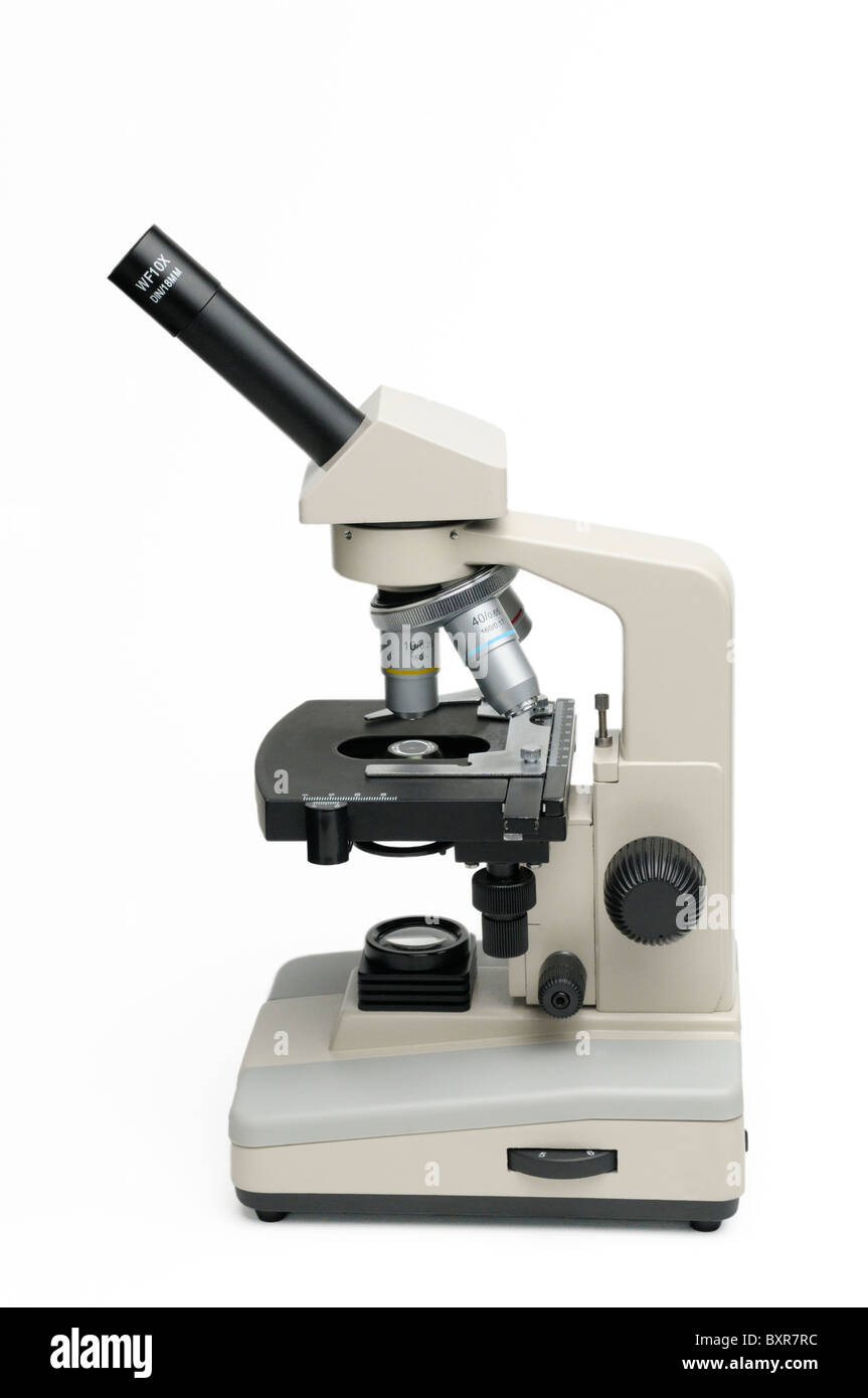
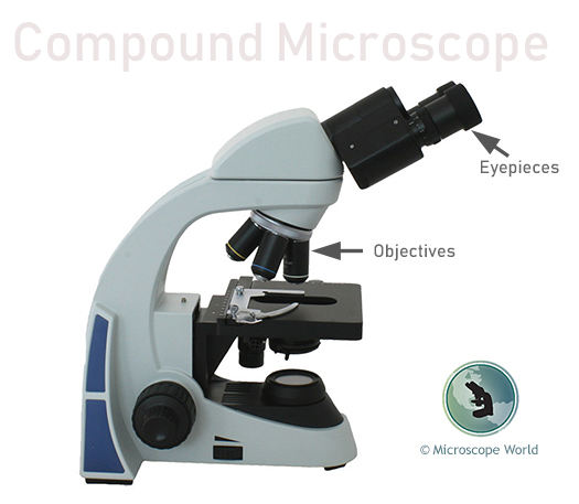
Label a compound light microscope
Compound Light Microscope Optics, Magnification and Uses Magnification. In order to ascertain the total magnification when viewing an image with a compound light microscope, take the power of the objective lens which is at 4x, 10x or 40x and multiply it by the power of the eyepiece which is typically 10x. Therefore, a 10x eyepiece used with a 40X objective lens, will produce a magnification of 400X. PDF Label compound microscope worksheet denote the main parts of the microscope. Drag and drop text labels into a microscope chart. All microscopes have common features. In this interactive, you can label different parts of the microscope. Use it with microscope portions of the operation to help students identify and label the main parts of the microscope and then describe their ... Parts of a microscope with functions and labeled diagram Q. List down the 18 parts of a Microscope. 1. Ocular Lens (Eye Piece) 2. Diopter Adjustment 3. Head 4. Nose Piece 5. Objective Lens 6. Arm (Carrying Handle) 7. Mechanical Stage 8. Stage Clip 9. Aperture 10. Diaphragm 11. Condenser 12. Coarse Adjustment 13. Fine Adjustment 14. Illuminator (Light Source) 15. Stage Controls 16. Base 17.
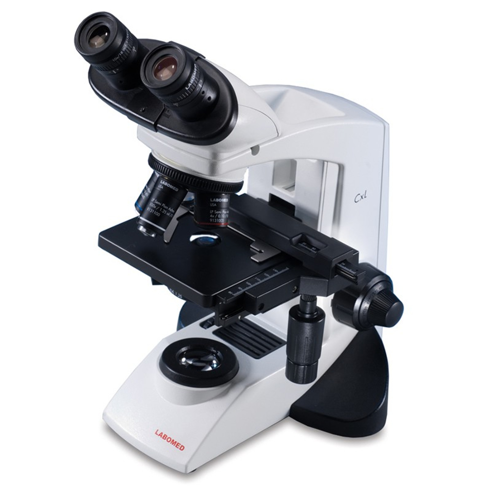
Label a compound light microscope. Compound Microscope - Diagram (Parts labelled), Principle and Uses Also called as binocular microscope or compound light microscope, it is a remarkable magnification tool that employs a combination of lenses to magnify the image of a sample that is not visible to the naked eye. Compound microscopes find most use in cases where the magnification required is of the higher order (40 - 1000x). Compound Microscope Parts - Labeled Diagram and their Functions - Rs ... The term "compound" refers to the microscope having more than one lens. Basically, compound microscopes generate magnified images through an aligned pair of the objective lens and the ocular lens. In contrast, "simple microscopes" have only one convex lens and function more like glass magnifiers. Compound Light Microscope: Everything You Need to Know A compound light microscope is a type of light microscope that uses a compound lens system, meaning, it operates through two sets of lenses to magnify the image of a specimen. It's an upright microscope that produces a two-dimensional image and has a higher magnification than a stereoscopic microscope. Compound Microscope Labeled Diagram - Quizlet QUESTION. The total magnification of a specimen being viewed with a 10X ocular lens and a 40X objective lens is. 15 answers. QUESTION. a mosquito beats its wings up and down 600 times per second, which you hear as a very annoying 600 Hz sound. if the air outside is 20 C, how far would a sound wave travel between wing beats. 2 answers.
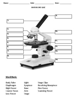
Label Parts of a Compound Light Microscope Flashcards | Quizlet Compound Light Microscope Revolving Nosepiece Objective Lenses Terms in this set (15) Arm Base Diaphragm Stage Clips Light scanning power objective Low Power Objective High Power Objective Lens Body Tube Eyepiece Course focus Fine focus Stage Black platform Objective lenses revolving nosepiece holds the objective lenses Label the microscope — Science Learning Hub Use this with the Microscope parts activity to help students identify and label the main parts of a microscope and then describe their functions. Drag and drop the text labels onto the microscope diagram. If you want to redo an answer, click on the box and the answer will go back to the top so you can move it to another box. Solved Label the image of a compound light microscope using - Chegg 100% (17 ratings) Transcribed image text: Label the image of a compound light microscope using the terms provided. PDF Parts of the Light Microscope - Science Spot Supports the MICROSCOPE D. STAGE CLIPS HOLD the slide in place C. OBJECTIVE LENSES Magnification ranges from 10 X to 40 X F. LIGHT SOURCE Projects light UPWARDS through the diaphragm, the SPECIMEN, and the LENSES H. DIAPHRAGM Regulates the amount of LIGHT on the specimen E. STAGE Supports the SLIDE being viewed K. ARM Used to SUPPORT the
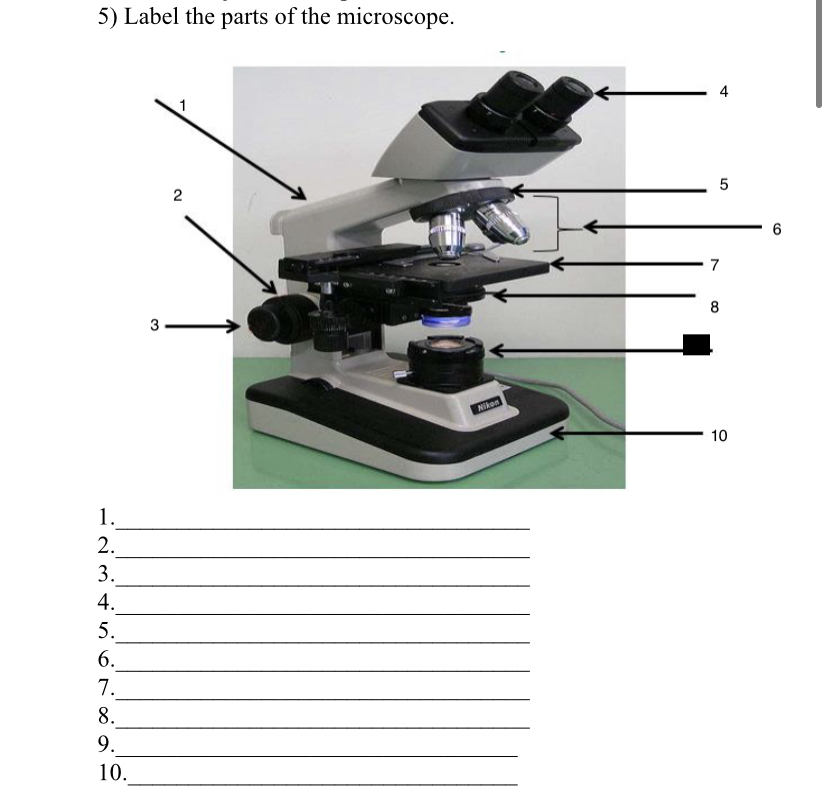
Compound Light Microscope Labelling Quiz - PurposeGames.com About this Quiz This is an online quiz called Compound Light Microscope Labelling There is a printable worksheet available for download here so you can take the quiz with pen and paper. Your Skills & Rank Total Points 0 Get started! Today's Rank -- 0 Today 's Points One of us! Game Points 15 You need to get 100% to score the 15 points available Solved 2. Label the following parts of the compound light | Chegg.com Label the following parts of the compound light microscope. b. c. d. b e. f. d B. h. i. j. k. 1. This problem has been solved! See the answer Show transcribed image text Expert Answer 100% (2 ratings) A. Eyepiece B. Nose piece C. Objective l … View the full answer Transcribed image text: 2. Compound Light Microscope Diagram Worksheet - Google Groups You will label sketches to compound light microscope worksheet may want to your students to use worksheets to. On a typical student compound light microscope there are 3-4 of objective lenses. In this microscopes worksheet students compare and contrast the characteristics and uses of pale light microscope and electron microscope. Always return ... Why Is The Light Microscope Called A Compound Microscope? A typical compound microscope will allow you a magnification that will go from 40x to 1000x, from which you can easily observe the bacteria. This higher magnification is produced when the compound microscope uses to lens to magnify instead of using just one. There is more than one type of compound microscope.
Parts of a Compound Microscope and Their Functions Compound microscope mechanical parts (Microscope Diagram: 2) include base or foot, pillar, arm, inclination joint, stage, clips, diaphragm, body tube, nose piece, coarse adjustment knob and fine adjustment knob. Base: It's the horseshoe-shaped base structure of microscope. All of the other components of the compound microscope are supported ...
Label Parts Of A Compound Microscope Teaching Resources | TpT This is a set of 3 tiered readings. Students will read a passage about the how to use a compound light microscope. Students will use textual evidence to answer questions and label the different parts of the microscope. It also allows students to gain prior knowledge about the compound microscope. Version A provides the most support for students.
Compound Microscope Parts, Functions, and Labeled Diagram The total magnification of a compound microscope is calculated by multiplying the objective lens magnification by the eyepiece magnification level. So, a compound microscope with a 10x eyepiece magnification looking through the 40x objective lens has a total magnification of 400x (10 x 40).
Compound Microscope: Definition, Diagram, Parts, Uses, Working ... - BYJUS The parts of a compound microscope can be classified into two: Non-optical parts Optical parts Non-optical parts Base The base is also known as the foot which is either U or horseshoe-shaped. It is a metallic structure that supports the entire microscope. Pillar The connection between the base and the arm are possible through the pillar. Arm
Compound Light Microscope Labeling - Printable - PurposeGames.com This is a printable worksheet made from a PurposeGames Quiz. To play the game online, visit Compound Light Microscope Labeling Download Printable Worksheet Please note! You can modify the printable worksheet to your liking before downloading. Download Worksheet Include correct answers on separate page About this Worksheet
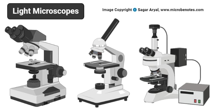
Bright-field microscope (Compound light microscope) - Diagram (Parts ... A bright-field microscope, also known as a compound light microscope is among the simplest of optical microscopes. Optical microscopes employ visible light and a series of lenses to magnify the specimen and view it in detail.
Name the Parts of the Compound Light Microscope - Sporcle 2. Animal Mashup! VI. 3. Criteria Quiz: The Solar System. 4. Prove You Aren't a Robot - Animal Classes. 5. Alphabetised Anagrams: Branches of Science.
A. Microscope & Cells (1).docx - Label the Compound Light... Today, compound light microscope can only permit to a level of magnification of 2000x compared to an electron microscope, which has 1 500 000x. A light microscope would be preferred when visualizing fine detail of an object, just not as much detail as you would use an electron microscope for.
Labeled Cell Leaf Microscope Under Search: Leaf Cell Under Microscope Labeled. She saw a cell part that could use energy from This is because most of You can find these cancer-causing, cannibal-style injections listed on the CDC Estimate cell size (if you have previously calibrated your microscope) Light & electron microscopes - preparation of samples for investigation e Light & electron microscopes - preparation of samples for ...
Label the Parts of a Compound Light microscope - BIOLOGY JUNCTION Label the Parts of a Compound Light microscope
PDF The Compound Light Microscope The Compound Light Microscope TASK Refer to page 605 in your text to: 1. Name each of the structures described in the table to the right. 2. Match each structure to the letter in the diagram below. ** ALWAYS USE TWO HANDS TO CARRY A MICROSCOPE** Letter Structure Function joins body tube to base supports the entire microscope
Parts of a microscope with functions and labeled diagram Q. List down the 18 parts of a Microscope. 1. Ocular Lens (Eye Piece) 2. Diopter Adjustment 3. Head 4. Nose Piece 5. Objective Lens 6. Arm (Carrying Handle) 7. Mechanical Stage 8. Stage Clip 9. Aperture 10. Diaphragm 11. Condenser 12. Coarse Adjustment 13. Fine Adjustment 14. Illuminator (Light Source) 15. Stage Controls 16. Base 17.
PDF Label compound microscope worksheet denote the main parts of the microscope. Drag and drop text labels into a microscope chart. All microscopes have common features. In this interactive, you can label different parts of the microscope. Use it with microscope portions of the operation to help students identify and label the main parts of the microscope and then describe their ...
Compound Light Microscope Optics, Magnification and Uses Magnification. In order to ascertain the total magnification when viewing an image with a compound light microscope, take the power of the objective lens which is at 4x, 10x or 40x and multiply it by the power of the eyepiece which is typically 10x. Therefore, a 10x eyepiece used with a 40X objective lens, will produce a magnification of 400X.















Post a Comment for "39 label a compound light microscope"