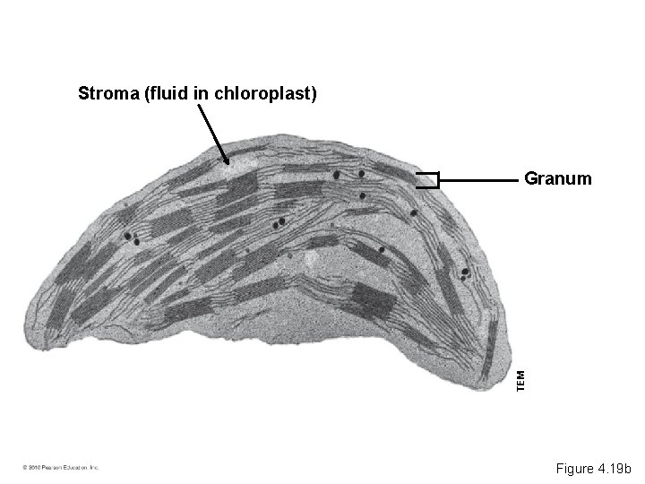
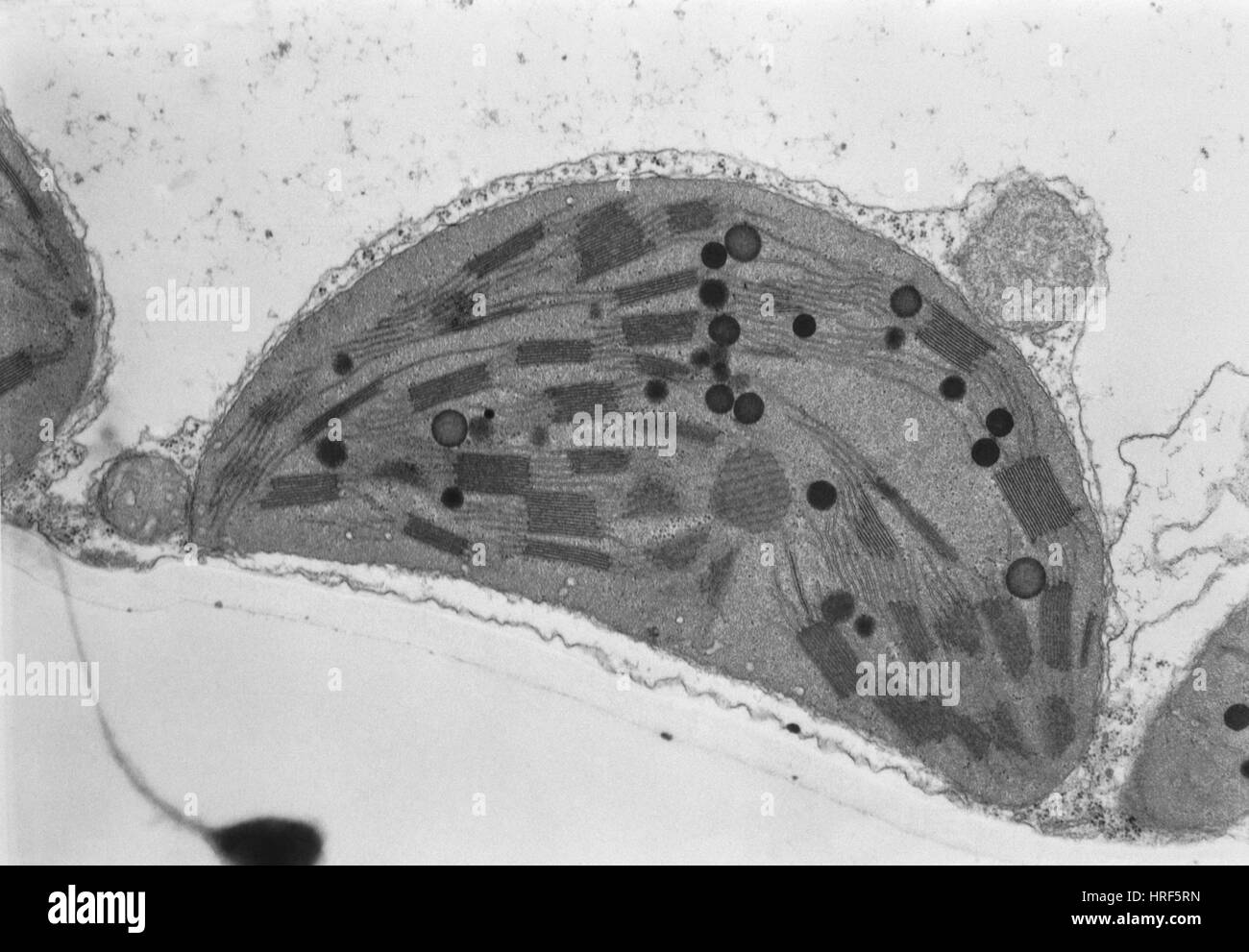
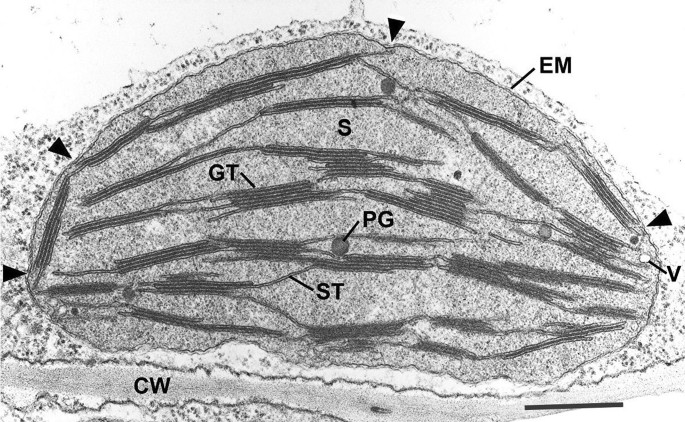
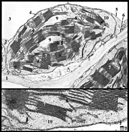
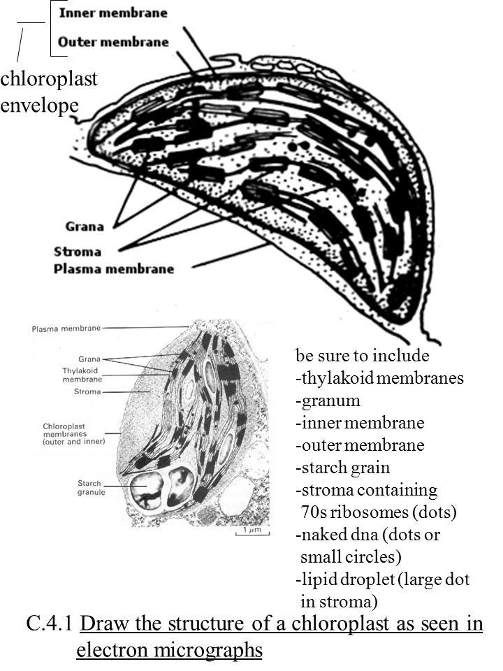
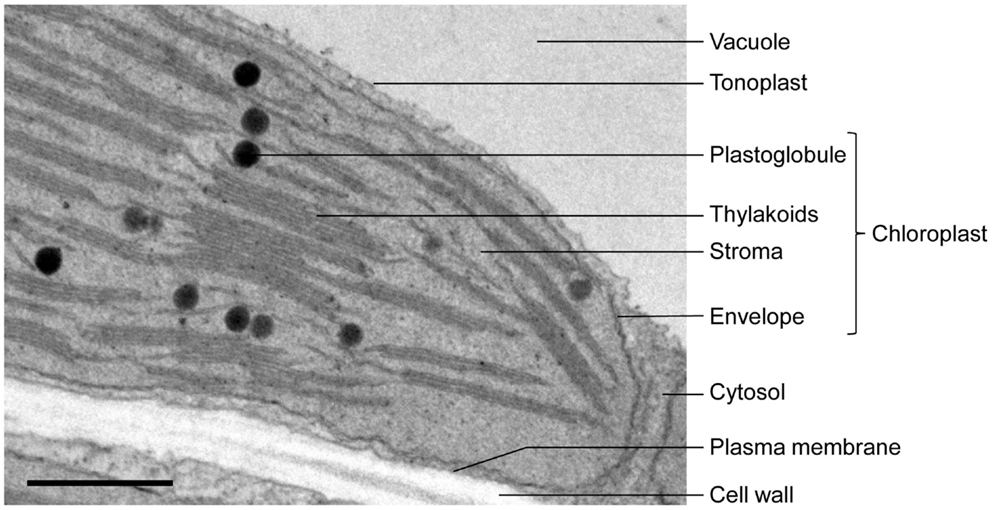
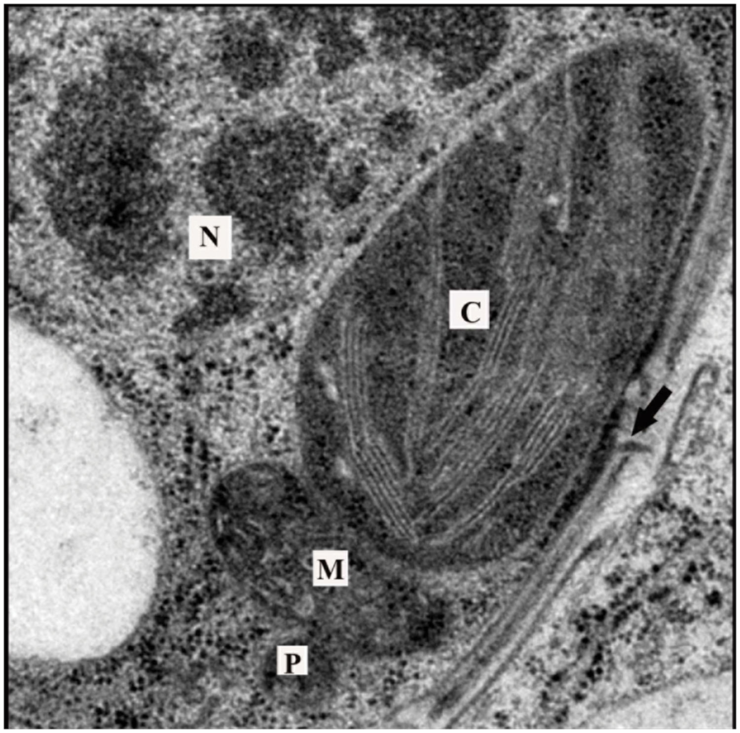
42 transmission electron microscope image of chloroplast labeled
Top 14 Types of Spectroscopic Techniques – Explained! ADVERTISEMENTS: Some of the important types of Spectroscopic Techniques are as follows: Type # 1. Gamma Spectroscopy: Gamma spectroscopy is a radionuclide measurement method. While a Geiger counter determines only the count rate, a gamma spectrometer will determine the energy and the count rate of gamma-rays emitted by radioactive substances. Gamma … The key micronutrient copper orchestrates broad-spectrum virus ... 1.7.2022 · Samples were observed by a field emission high resolution transmission electron microscope (JEM-2100F, Japan) at PKUAIC, Beijing, China. The regions of intercellular space, chloroplast, mitochondrion, nuclei, and intracellular space were visualized under TEM, and then the elemental contents in the selected area were measured by coupling EDS ( 46 ).
Scanning Electron Microscopic Study of Modified Chloroplasts ... by H Terai · 2000 · Cited by 13 — Scanning electron micrographs of various stages of chloroplasts in the sepal cells of broccoli florets during storage at 20 °C. In the preparation of specimens.

Transmission electron microscope image of chloroplast labeled
Amazing 27 Things Under The Microscope With Diagrams May 13, 2022 · A scanning transmission electron microscope (STEM) is often used to observe crystals or compounds that reveal the atoms present inside the compounds with some electrons being used to identify atoms of a particular element through the microscope. The structure of an atom is visible with these microscopes. Looking at the Structure of Cells in the Microscope A typical animal cell is 10–20 μm in diameter, which is about one-fifth the size of the smallest particle visible to the naked eye. It was not until good light microscopes became available in the early part of the nineteenth century that all plant and animal tissues were discovered to be aggregates of individual cells. This discovery, proposed as the cell doctrine by Schleiden and … Brassica napus BnaNTT1 modulates ATP homeostasis in Jul 12, 2022 · Then, the chloroplast ultrastructure of mesophyll cells in leaves was observed under a transmission electron microscope. c6-1b a7-1a mutants exhibited smaller chloroplast size and impaired thylakoid structure with a smaller number and irregular distribution of grana, while larger starch particles appeared in the chloroplasts in OE leaves.
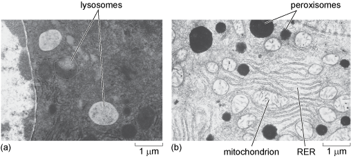
Transmission electron microscope image of chloroplast labeled. Transmission Electron Micrograph Of A Chloroplast ... - Getty Images View top-quality stock photos of Transmission Electron Micrograph Of A Chloroplast. Find premium, high-resolution stock photography at Getty Images. Micro Exam 1 Study Questions Flashcards | Quizlet Which of the following microscopes is capable of producing a three-dimensional image? A. Differential interference contrast B. Scanning electron ... _____ involves the application of labeled antibodies to the specimen that is to be observed. a ... Transmission electron microscope. d. Preparing a specimen for TEM involves all of the following ... ESSENTIAL CELL BIOLOGY ESSENTIAL CELL BIOLOGY Academia.edu is a platform for academics to share research papers. Ribosome – protein factory – definition, function, structure, and ... Instead, a transmission electron microscope (TEM) is required to view ribosomes and rough endoplasmic reticulum. References “Ribosome” “Ribosome” by British Society for Cell Biology “ Nucleus and ribosomes ” “Structure and Function of the Eukaryotic Ribosome” by Jennifer A Doudna and Virginia L Rath “Ribosome database project”
Defects in autophagy lead to selective in vivo changes in turnover … Jun 29, 2022 · After several acetone rinses and the planchettes removed, samples were infiltrated in a series of Epon resin changes polymerizing at 60°C for 24 h. Sections were stained with 2% uranyl acetate and lead citrate (2.6% lead nitrate and 3.5% sodium citrate, pH 12) and imaged in a Philips CM120 transmission electron microscope. Ciliary transition zone proteins coordinate ciliary protein … 9.7.2022 · b Negative stain transmission electron micrographs of purified ciliary ectosomes (black arrows) from gametes in the mating process. 21gr (WT), ectosomes from mating supernatant of 21gr × 6145c ... Brassica napus BnaNTT1 modulates ATP homeostasis in Jul 12, 2022 · Then, the chloroplast ultrastructure of mesophyll cells in leaves was observed under a transmission electron microscope. c6-1b a7-1a mutants exhibited smaller chloroplast size and impaired thylakoid structure with a smaller number and irregular distribution of grana, while larger starch particles appeared in the chloroplasts in OE leaves. Looking at the Structure of Cells in the Microscope A typical animal cell is 10–20 μm in diameter, which is about one-fifth the size of the smallest particle visible to the naked eye. It was not until good light microscopes became available in the early part of the nineteenth century that all plant and animal tissues were discovered to be aggregates of individual cells. This discovery, proposed as the cell doctrine by Schleiden and …
Amazing 27 Things Under The Microscope With Diagrams May 13, 2022 · A scanning transmission electron microscope (STEM) is often used to observe crystals or compounds that reveal the atoms present inside the compounds with some electrons being used to identify atoms of a particular element through the microscope. The structure of an atom is visible with these microscopes.
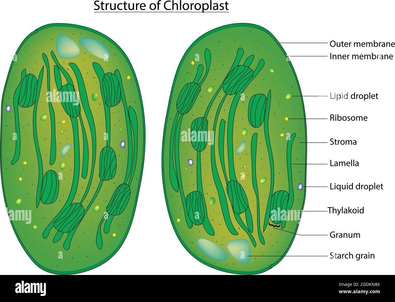
Cell theory, Plant cell diagram, Cell diagram

The plant-specific function of 2-Cys peroxiredoxin-mediated ...

Chloroplasts (13.1.1) | CIE A Level Biology Revision Notes ...

Draw a labelled diagram of a plant cell as revealed by an electron microscope. | 9 | IMPROVEMEN...

how to draw chloroplast | how to draw electron micrograph of chloroplast step by step

Chloroplast, TEM - Stock Image - C018/5179 - Science Photo ...

DP Topic 1.1 / 1.2 | Biology - Quizizz

1 Chloroplast. (a) Electron micrograph of a chloroplast in a ...

Transmission electron microscopic images of chloroplasts and ...

Warm Up 11/4/13 | Heena Bio HL YAY

TEM Chloroplast Labeling Diagram | Quizlet
A brief history of how microscopic studies led to the ...
A Thylakoid Membrane Protein Functions Synergistically with ...

Transmission electron microscopy picture of the chloroplast ...
Chloroplast - Wikipedia
Too rigid to fold: Carotenoid-dependent decrease in thylakoid ...
Chapter 4 A Tour of the Cell Power

Thin section electron micrograph of a chloroplast from wild ...
Chloroplast structure hi-res stock photography and images - Alamy
Chloroplast tem hi-res stock photography and images - Alamy
A brief history of how microscopic studies led to the ...
A tour of the cell: View as single page

Electron Microscopy Cells Photos - Free & Royalty-Free Stock ...
Leaf chloroplast
2.2.1 Draw a generalized prokaryotic cell as seen in electron ...

TEM of chloroplast from Coleus blumei - Stock Image - B110 ...
Frontiers | When Proteomics Reveals Unsuspected Roles: The ...

Transmission electron micrograph of chloroplast in Scots pine ...

File:Chloroplast in leaf of Anemone sp TEM 30000x.png ...
Preparation of plant cells for transmission electron ...
A JEOL JEM1010TEM Transmission Electron Microscope ...
A. Electron microscopy picture of a higher plant chloroplast ...

Pin page
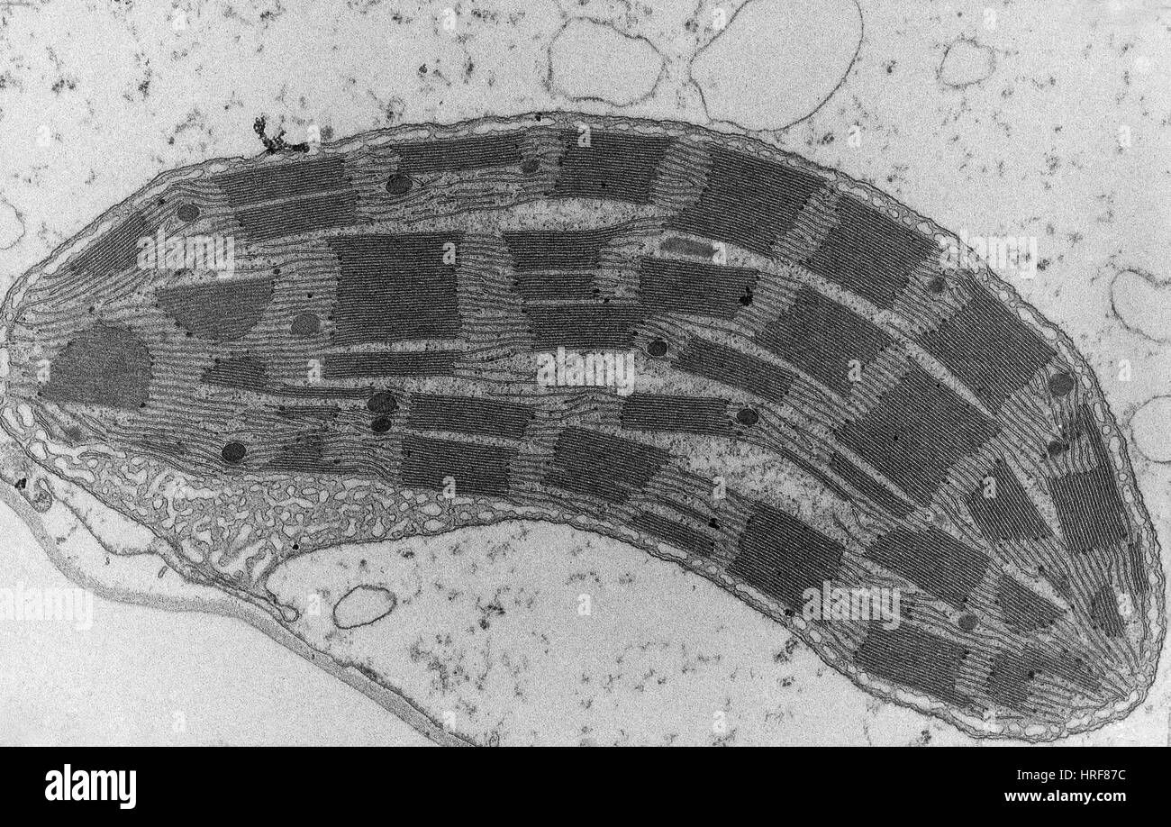
Chloroplast tem hi-res stock photography and images - Alamy
Frontiers | Chloroplast signaling within, between and beyond ...

Electron micrograph of an Arabidopsis leaf chloroplast ...

Thin section electron micrograph of a young tobacco ...
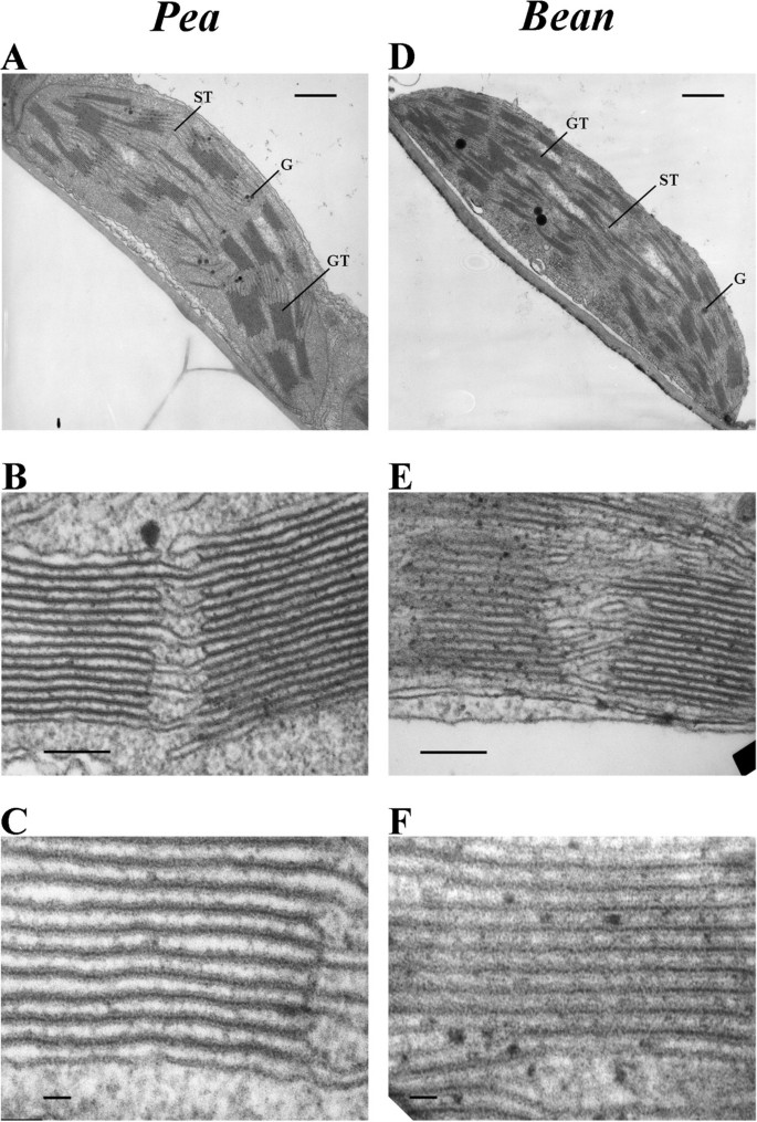
Correlation between spatial (3D) structure of pea and bean ...

Transmission electron microscopy (TEM) observation of ...
Histopathology of Chloroplast Ultrastructure and Inclusion ...
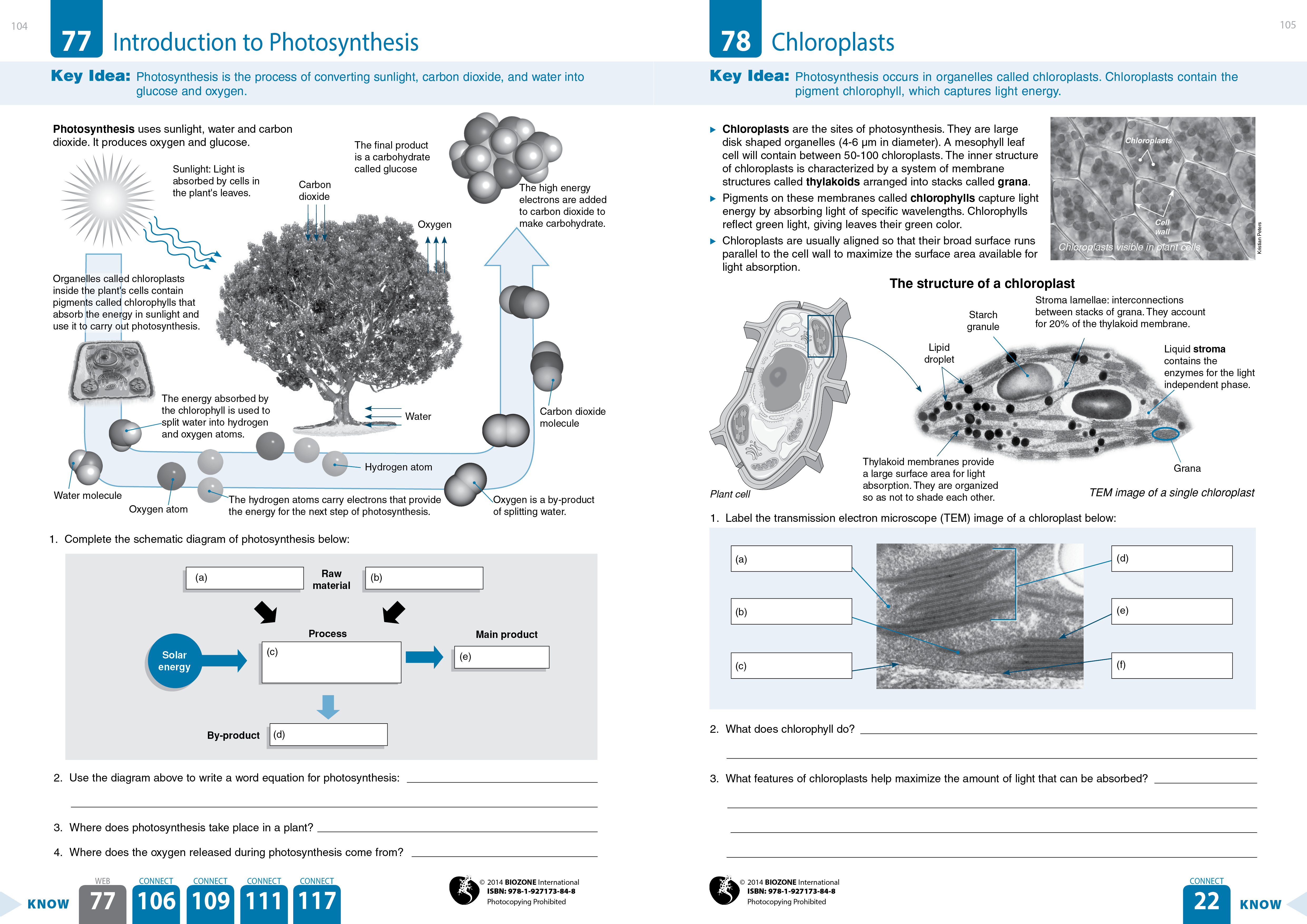
30 Label The Transmission Electron Microscope Image Of A ...

TEM of chloroplasts in a leaf cell - Stock Image - B110/0033 ...
Post a Comment for "42 transmission electron microscope image of chloroplast labeled"