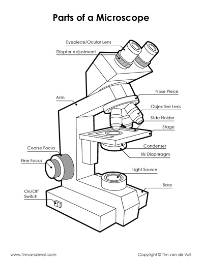
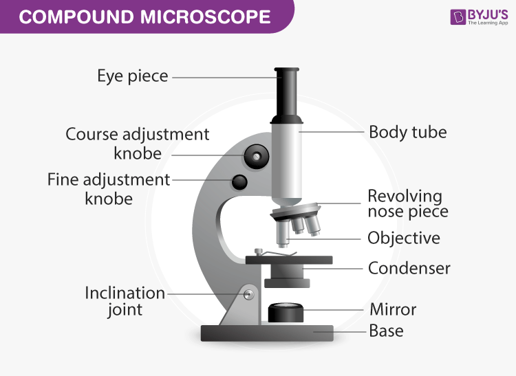
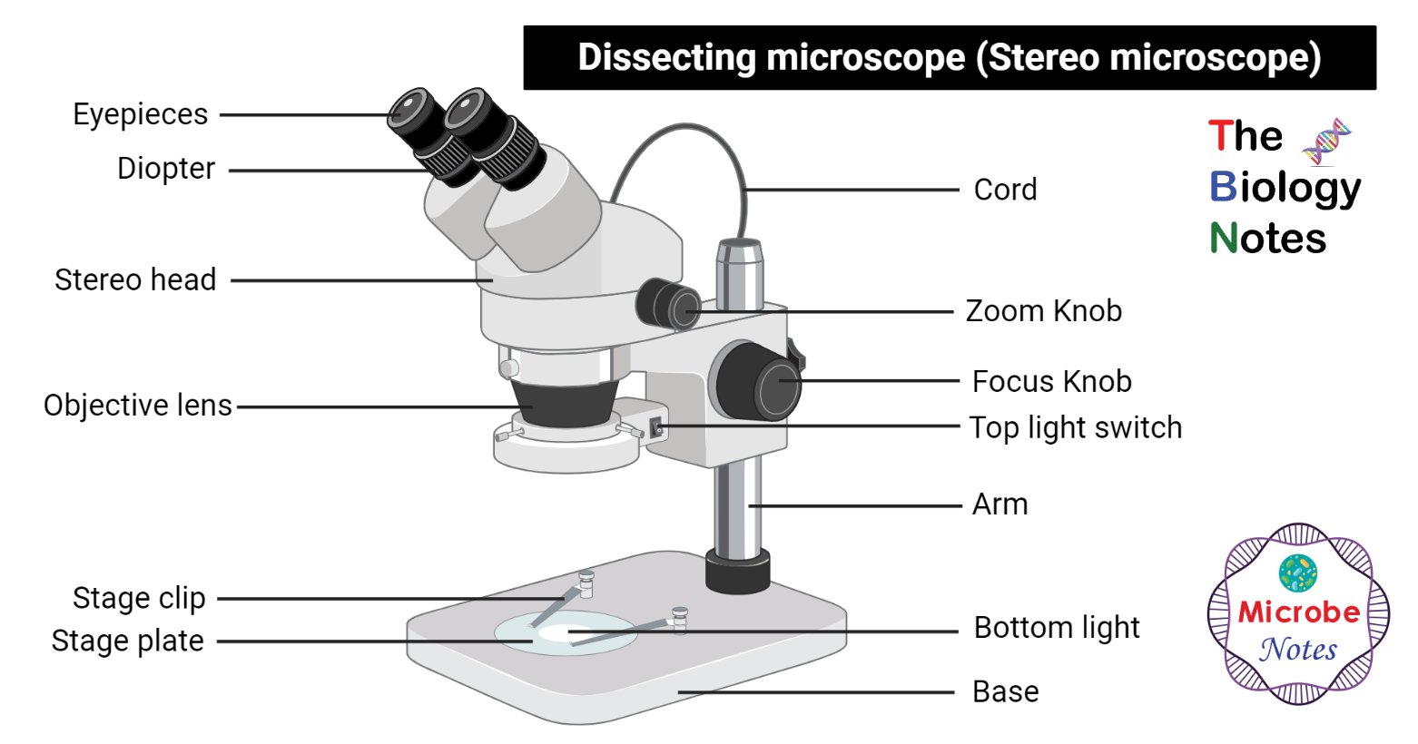
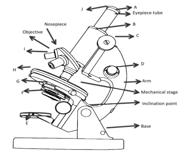
43 diagram of a microscope with labels
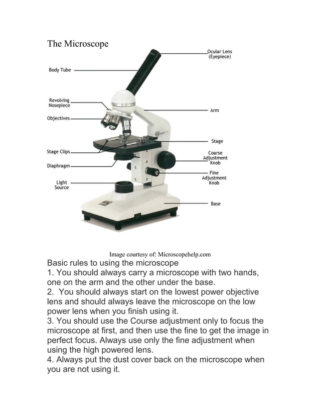
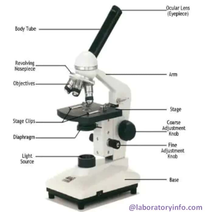
Parts of a microscope with functions and labeled diagram - Microbe Notes Parts of a microscope with functions and labeled diagram September 17, 2022 by Faith Mokobi Having been constructed in the 16th Century, Microscopes have revolutionalized science with their ability to magnify small objects such as microbial cells, producing images with definitive structures that are identifiable and characterizable. Label Microscope Diagram - EnchantedLearning.com Label Microscope Diagram Using the terms listed below, label the microscope diagram. Inventions and Inventors arm - this attaches the eyepiece and body tube to the base. base - this supports the microscope. body tube - the tube that supports the eyepiece. coarse focus adjustment - a knob that makes large adjustments to the focus.
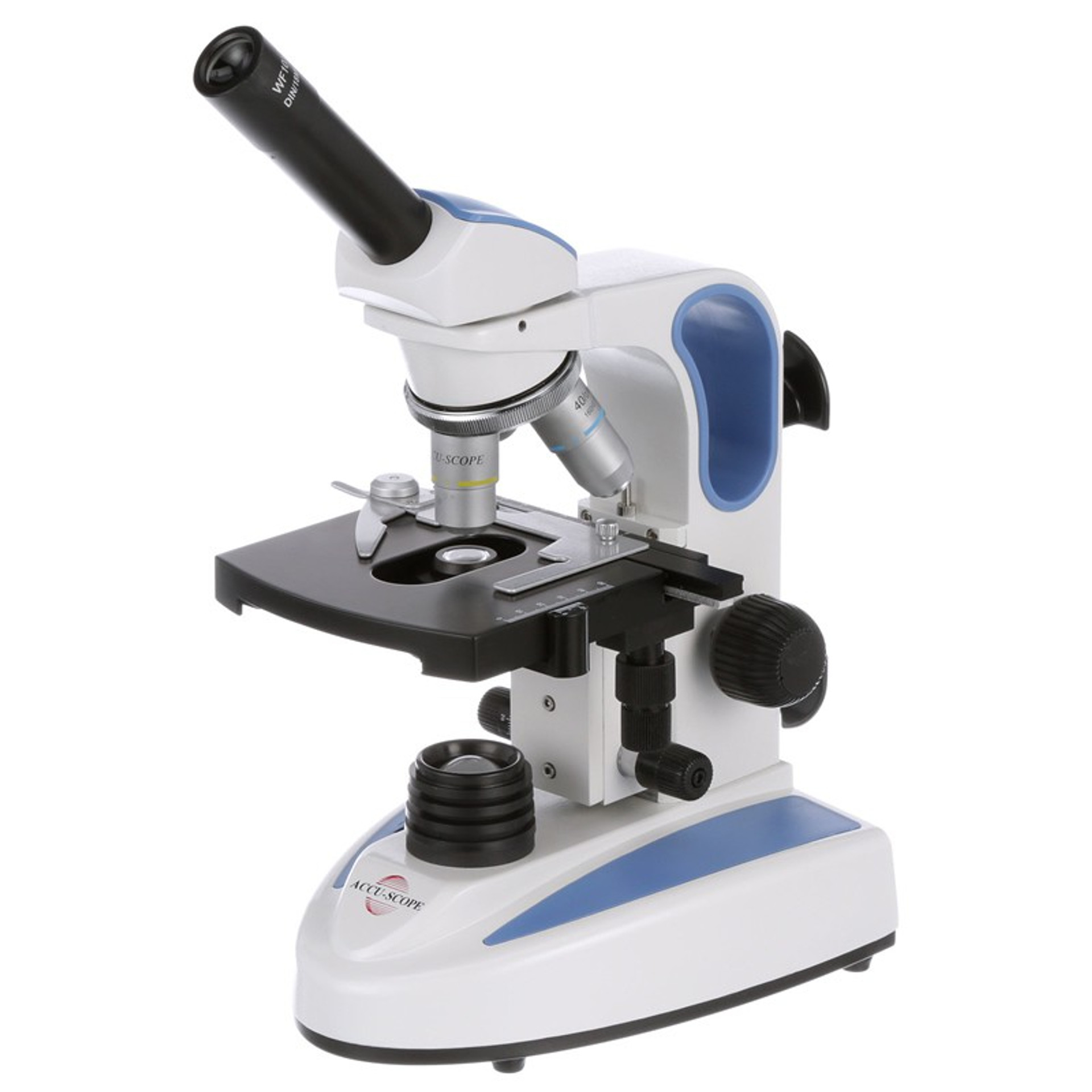
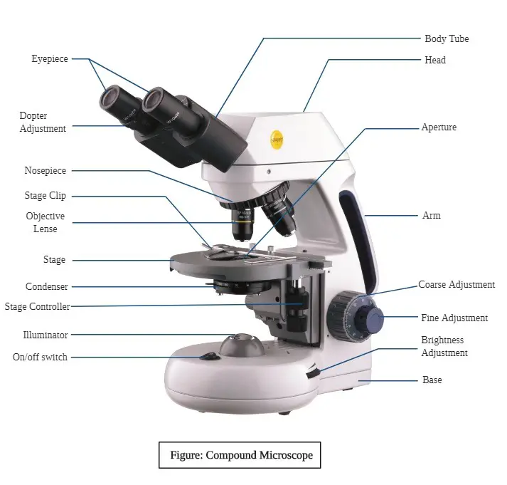
Compound Microscope Parts, Functions, and Labeled Diagram Compound Microscope Definitions for Labels Eyepiece (ocular lens) with or without Pointer: The part that is looked through at the top of the compound microscope. Eyepieces typically have a magnification between 5x & 30x. Monocular or Binocular Head: Structural support that holds & connects the eyepieces to the objective lenses.
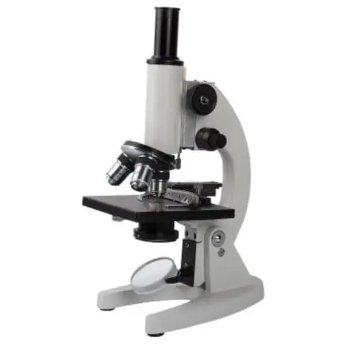
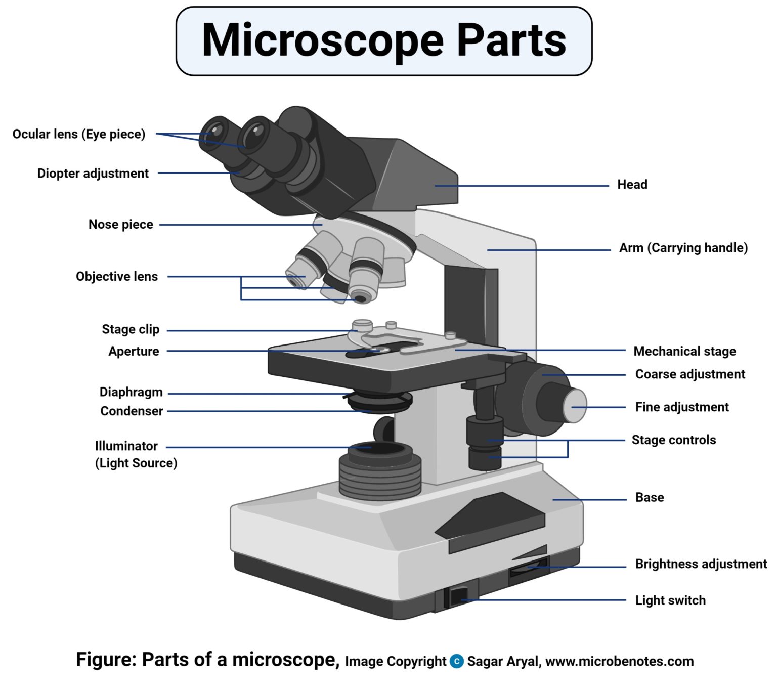
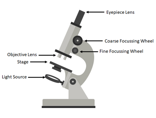
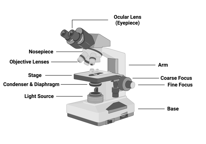
Diagram of a microscope with labels
Tongue Under Microscope with Labeled Diagram - AnatomyLearner This dog tongue labeled diagram also shows the lamina propria and skeletal muscle bundles. The ruminant tongue labeled diagram shows the vallate papillae with a surrounding sulcus and taste buds. You will find more labeled diagrams on the tongue of different species like rabbits, pigs, and lions on the social media of anatomy learners. Conclusion › pages › examplesDiagram Maker - Free Online Diagram Templates | Lucidchart A diagram is a symbolic representation of information that helps you visualize concepts. It shows the arrangement of ideas or elements and how they relate to one another. Today, you’ll find diagrams in numerous fields, including education, writing, engineering, and marketing. 16 Parts of a Compound Microscope: Diagrams and Video The 16 core parts of a compound microscope are: Head (Body) Arm Base Eyepiece Eyepiece tube Objective lenses Revolving Nosepiece (Turret) Rack stop Coarse adjustment knobs Fine adjustment knobs Stage Stage clips Aperture Illuminator Condenser Diaphragm Video: Parts of a compound Microscope with Diagram Explained
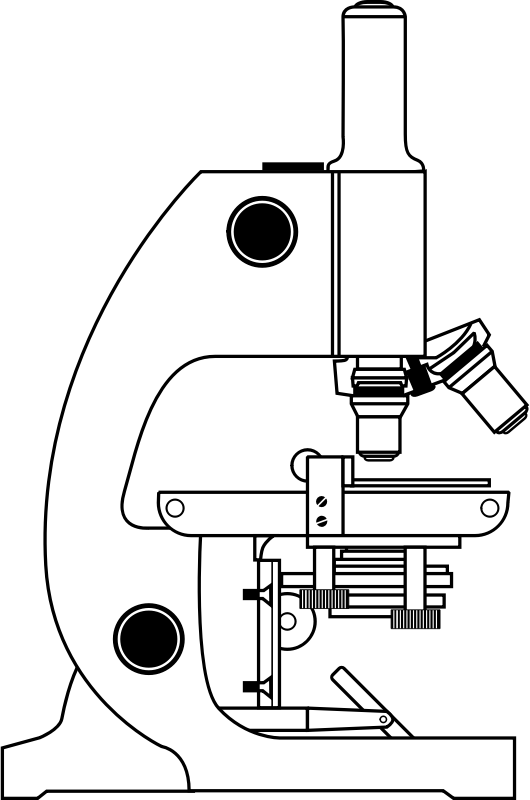
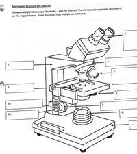
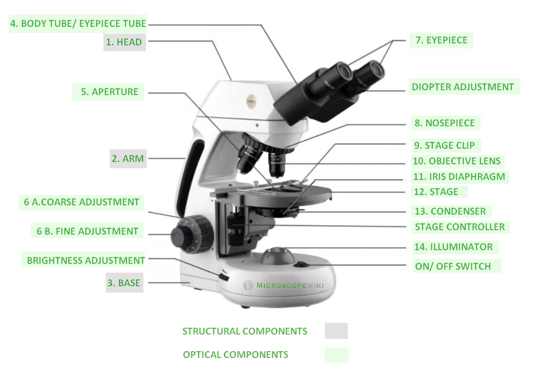
Diagram of a microscope with labels. Microscope Labeling - The Biology Corner Microscope Labeling. This simple worksheet pairs with a lesson on the light microscope, where beginning biology students learn the parts of the light microscope and the steps needed to focus a slide under high power. The labeling worksheet could be used as a quiz or as part of direct instruction where students label the microscope as you go ... Parts of the Microscope with Labeling (also Free Printouts) Parts of the Microscope with Labeling (also Free Printouts) By Editorial Team March 7, 2022 A microscope is one of the invaluable tools in the laboratory setting. It is used to observe things that cannot be seen by the naked eye. Table of Contents 1. Eyepiece 2. Body tube/Head 3. Turret/Nose piece 4. Objective lenses 5. Knobs (fine and coarse) 6. Compound Microscope - Diagram (Parts labelled), Principle and Uses See: Labeled Diagram showing differences between compound and simple microscope parts. Structural Components. The three structural components include. 1. Head. This is the upper part of the microscope that houses the optical parts. 2. Arm . This part connects the head with the base and provides stability to the microscope. Microscope Types (with labeled diagrams) and Functions Electron microscope labeled diagram The different types of electron microscopes are: Transmission Electron Microscope Scanning Electron Microscope Reflection Electron Microscope Scanning transmission electron microscope Scanning tunneling microscopy Electron microscope functions: Semiconductors and Data Storage Industry Failure Analysis
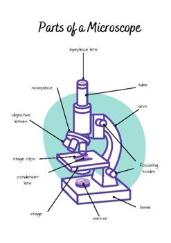
Parts of the Microscope (Labeled Diagrams) Simple microscope labelled diagram Image created with Biorender Tube/Body Tube It serves as the connector between the eyepiece/ocular and objective lenses. Objective lenses The lenses have varying magnifying power, which typically consists of 10x, 40x, and 100x. Microscope Parts, Types & Diagram | What is a Microscope? Microscope Diagram There are many illustrations available for the diagram of a light microscope. The essential parts include the head, base, arms, lenses, and lights. In diagrams, one... Labeling the Parts of the Microscope | Microscope World Resources Labeling the Parts of the Microscope This activity has been designed for use in homes and schools. Each microscope layout (both blank and the version with answers) are available as PDF downloads. You can view a more in-depth review of each part of the microscope here. Download the Label the Parts of the Microscope PDF printable version here. Microscope Parts and Functions 5 Hobby Microscopes for Beginners First, the purpose of a microscope is to magnify a small object or to magnify the fine details of a larger object in order to examine minute specimens that cannot be seen by the naked eye. Here are the important compound microscope parts... Eyepiece: The lens the viewer looks through to see the specimen.
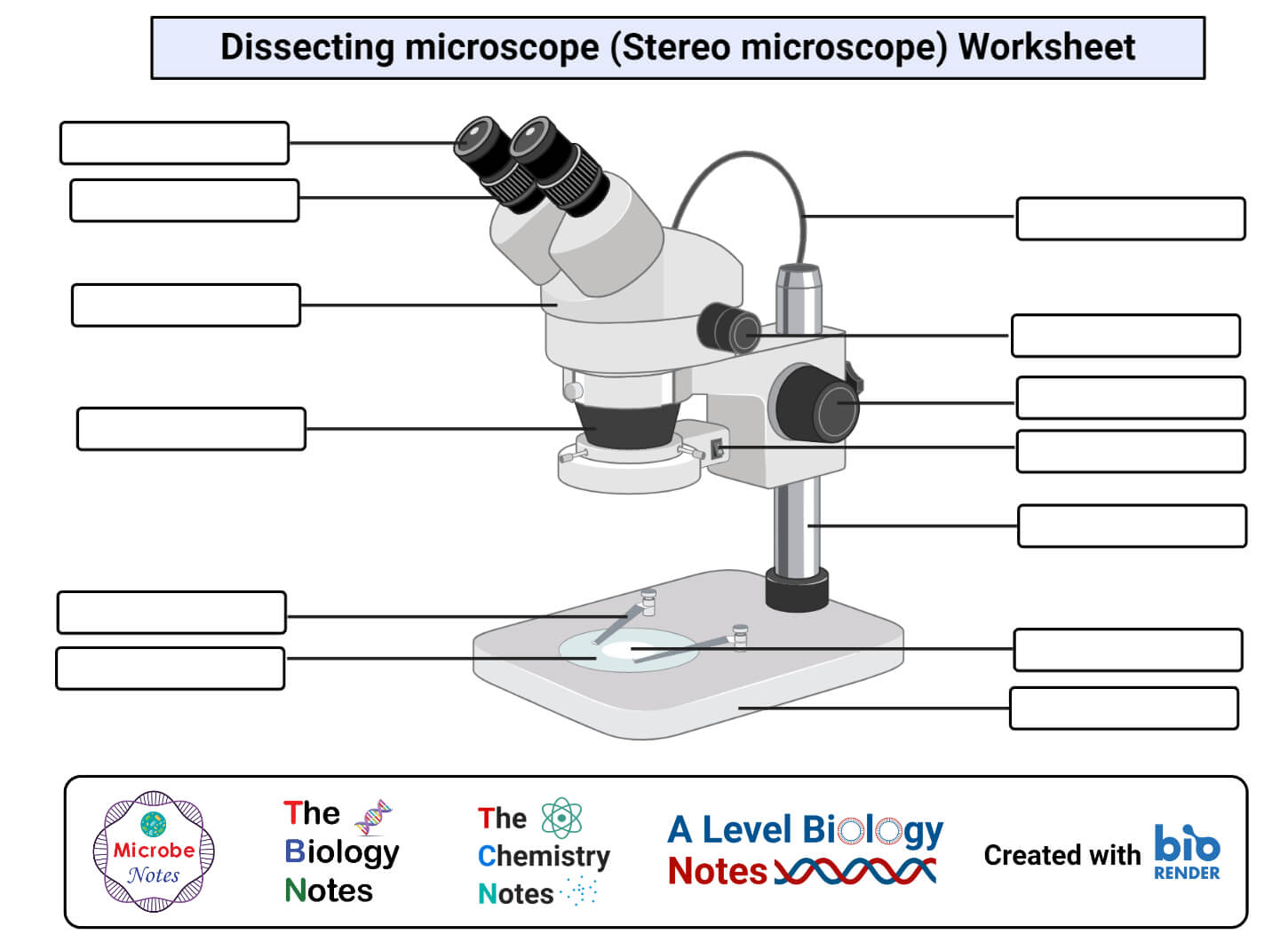
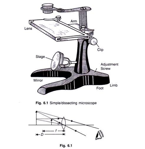
Parts of Stereo Microscope (Dissecting microscope) - labeled diagram ... Labeled part diagram of a stereo microscope Major structural parts of a stereo microscope. There are three major structural parts of a stereo microscope. The viewing Head includes the upper part of the microscope, which houses the most critical optical components, including the eyepiece, objective lens, and light source of the microscope. Compound light microscope diagram and functions. Microscope Parts and ... Compound Microscope Parts, Functions, and Labeled Diagram. Then the phase was raised up utilizing the harsh accommodation by detecting from the side until the slide is about touching the nonsubjective lens. They are usually stacked upon one another. ... Compound Microscope: Definition, Diagram, Parts, Uses, Working Principle. Simple Microscope - Diagram (Parts labelled), Principle, Formula and Uses Simple Microscope - Diagram (Parts labelled), Principle, Formula and Uses Simple Microscope By Editorial Board October 1, 2022 Dating back to the 14th century, simple microscope is the most basic of the various microscopes available. It is a type of optical microscope that uses visible light and lens to magnify objects. › diagramDiagram - definition of diagram by The Free Dictionary di·a·gram. (dī′ə-grăm′) n. 1. A plan, sketch, drawing, or outline designed to demonstrate or explain how something works or to clarify the relationship between the parts of a whole. 2. Mathematics A graphic representation of an algebraic or geometric relationship. 3. A chart or graph.
How to draw compound of Microscope easily - step by step I will show you " How to draw compound of microscope easily - step by step "Please watch carefully and try this okay.Thanks for watching.....#microscopedrawi...
Microscope Parts, Function, & Labeled Diagram - slidingmotion Microscope Parts Labeled Diagram The principle of the Microscope gives you an exact reason to use it. It works on the 3 principles. Magnification Resolving Power Numerical Aperture. Parts of Microscope Head Base Arm Eyepiece Lens Eyepiece Tube Objective Lenses Nose Piece Adjustment Knobs Stage Aperture Microscopic Illuminator Condenser Lens
Microscope: Parts Of A Microscope With Functions And Labeled Diagram. Figure: A diagram of a microscope's components The microscope has three basic components: the head, the base, and the arm. Head:Occasionally, the head is considered the body. It holds the optical components of the upper part of the microscope. Base:The microscope's base provides great support. It is also equipped with miniature illuminators.
PDF Label parts of the Microscope: Answers Label parts of the Microscope: Answers Coarse Focus Fine Focus Eyepiece Arm Rack Stop Stage Clip . Created Date: 20150715115425Z ...
Microscope Labeling Diagram | Quizlet Coarse Focus Knob Moves the stage large distances to roughly focus the image. Fine Focus Knob Moves the stage tiny distances to slightly adjust and fine-tune the image focus. Arm Supports the body tube. Objective Lenses Focus and magnify light in differing amounts to view the specimen. Stage Clips Hold the slide in place on the stage. Nosepiece
Label the microscope — Science Learning Hub All microscopes share features in common. In this interactive, you can label the different parts of a microscope. Use this with the Microscope parts activity to help students identify and label the main parts of a microscope and then describe their functions. Drag and drop the text labels onto the microscope diagram.
Microscope, Microscope Parts, Labeled Diagram, and Functions The description given below summarize the brief description of microscope parts used to visualize the microscopic specimens such as animal cells, plant cells, microbes, bacteria, viruses, microorganisms etc. The Microscopes parts divided into three different structural parts Head, Base, and Arms.
Compound Microscope Parts - Labeled Diagram and their Functions Labeled diagram of a compound microscope Major structural parts of a compound microscope Optical components of a compound microscope Eyepiece Eyepiece tube Objective lenses Nosepiece Specimen stage Coarse and fine focus knobs Rack stop Illuminator Condenser Abbe condenser Iris Diaphragm Condenser Focus Knob Summary An overview of microscopes
› blog › how-to-draw-architectural-diagramsHow to draw 5 types of architectural diagrams - Lucidchart An architectural diagram is a visual representation that maps out the physical implementation for components of a software system. It shows the general structure of the software system and the associations, limitations, and boundaries between each element. Software environments are complex—and they aren’t static.
A Study of the Microscope and its Functions With a Labeled Diagram ... A Study of the Microscope and its Functions With a Labeled Diagram To better understand the structure and function of a microscope, we need to take a look at the labeled microscope diagrams of the compound and electron microscope. These diagrams clearly explain the functioning of the microscopes along with their respective parts.
› browse › diagramDiagram Definition & Meaning | Dictionary.com noun. a figure, usually consisting of a line drawing, made to accompany and illustrate a geometrical theorem, mathematical demonstration, etc. a drawing or plan that outlines and explains the parts, operation, etc., of something: a diagram of an engine. a chart, plan, or scheme.
Simple Microscope - Parts, Functions, Diagram and Labelling Parts of the optical parts are as follows: Mirror - A simple microscope has a plano-convex mirror and its primary function is to focus the surrounding light on the object being examined. Lens - The biconvex lens is placed above the stage and its function is to magnify the size of the object being examined.
Compound Microscope: Definition, Diagram, Parts, Uses, Working ... - BYJUS The parts of a compound microscope can be classified into two: Non-optical parts Optical parts Non-optical parts Base The base is also known as the foot which is either U or horseshoe-shaped. It is a metallic structure that supports the entire microscope. Pillar The connection between the base and the arm are possible through the pillar. Arm
support.microsoft.com › en-us › officeCreate a Venn diagram - Microsoft Support Overview of Venn diagrams. A Venn diagram uses overlapping circles to illustrate the similarities, differences, and relationships between concepts, ideas, categories, or groups. Similarities between groups are represented in the overlapping portions of the circles, while differences are represented in the non-overlapping portions of the circles. 1 Each large group is represented by one of the circles.
› thesaurus › diagram97 Synonyms & Antonyms of DIAGRAM - Merriam-Webster Synonyms for DIAGRAM: illustration, graphic, drawing, picture, visual, image, figure, caption; Antonyms of DIAGRAM: integrate, consolidate, synthesize, unify, aggregate, conglomerate, coalesce, assimilate
Light microscopes - Cell structure - Edexcel - BBC Bitesize The components of a light microscope and their functions Calculating the magnification of light microscopes. The compound microscope uses two lenses to magnify the specimen: the eyepiece and an ...
Labelled Diagram of Compound Microscope The below mentioned article provides a labelled diagram of compound microscope. Part # 1. The Stand: The stand is made up of a heavy foot which carries a curved inclinable limb or arm bearing the body tube. The foot is generally horse shoe-shaped structure (Fig. 2) which rests on table top or any other surface on which the microscope in kept.
diagram.comDiagram Design Smarter. Magical new ways to design products. Automator. Automate your Figma tasks in one click. Magician. A magical design tool powered by AI. Learn more
16 Parts of a Compound Microscope: Diagrams and Video The 16 core parts of a compound microscope are: Head (Body) Arm Base Eyepiece Eyepiece tube Objective lenses Revolving Nosepiece (Turret) Rack stop Coarse adjustment knobs Fine adjustment knobs Stage Stage clips Aperture Illuminator Condenser Diaphragm Video: Parts of a compound Microscope with Diagram Explained
› pages › examplesDiagram Maker - Free Online Diagram Templates | Lucidchart A diagram is a symbolic representation of information that helps you visualize concepts. It shows the arrangement of ideas or elements and how they relate to one another. Today, you’ll find diagrams in numerous fields, including education, writing, engineering, and marketing.
Tongue Under Microscope with Labeled Diagram - AnatomyLearner This dog tongue labeled diagram also shows the lamina propria and skeletal muscle bundles. The ruminant tongue labeled diagram shows the vallate papillae with a surrounding sulcus and taste buds. You will find more labeled diagrams on the tongue of different species like rabbits, pigs, and lions on the social media of anatomy learners. Conclusion














Post a Comment for "43 diagram of a microscope with labels"